Immunoassays Compared for Diagnosis of Acute Murine Typhus Infections
|
By LabMedica International staff writers Posted on 10 Sep 2019 |
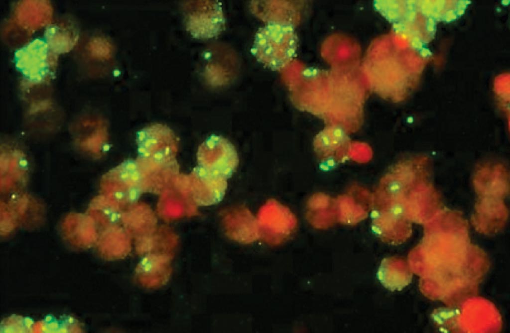
Image: A photomicrograph of an immunofluorescence assay (IFA) for Rickettsia (Photo courtesy of Fuller Laboratories).
Murine typhus is a disease transmitted by fleas and is caused by intracellular gram-negative bacteria called Rickettsia typhi, and manifested clinically with acute fever, chills, muscle pain, nausea, vomiting, stomach pain, cough and rash.
Appropriate rapid diagnostics are needed to distinguish it from other infections, as patient management varies. Due to low rickettsemia during acute illness, the sensitivity of real-time polymerase chain reaction (PCR) is highly variable. Thus, sero-diagnosis using immunofluorescence assay (IFA) remains the gold standard.
Scientists working with the Indonesia Research Partnership on Infectious Disease (Jakarta, Indonesia) obtained samples from eight government referral teaching hospitals in seven provincial capitals. Paired acute and convalescent plasma samples from 40 cases with confirmed R. typhi and 58 controls with another confirmed infection were used to evaluate the performance of commercial IgM and IgG enzyme-linked immunosorbent assay (ELISA) and IFA. The 58 paired plasma specimens that they used for controls were negative for R. typhi and Rickettsia spp., but positive for other pathogens by culture or molecular testing.
The immunofluorescence assay (IFA) was performed using kits from Focus Diagnostics (Cypress, CA, USA). The dilution for study samples was 1:64, and for provided positive controls was 1:32. Acute and convalescent specimens from each subject were performed simultaneously. Acute and convalescent plasma were tested simultaneously using ELISA kits from Fuller Laboratories (Fullerton, CA, USA). Microwells were coated with the outer surface membrane protein rOmp B purified from R. typhi. Optical density (OD) was measured at a wavelength of 450 nm.
The team reported that sensitivity and specificity of combined ELISA IgM and IgG anti-R. typhi using paired specimens were excellent (95.0% and 98.3%, respectively), comparable to combined IFA IgM and IgG (97.5% and 100%, respectively); sensitivity of ELISA IgM from acute specimens only was poor (45.0%), but specificity was excellent (98.3%). IFA IgM was more sensitive (77.5%), but less specific (89.7%) for single specimens. IgM was detected as early as day three of fever by ELISA and day four by IFA. Starting from day nine of illness, IgM was detected in all cases by IFA, while ELISA missed two specimens (days 10 and 25).
The authors concluded that their data supports the validity of ELISA in the diagnosis of R. typhi infection. As the specificity in acute specimens as well as sensitivity and specificity in convalescent specimens and paired specimens were excellent, ELISA is recommended when fluorescence microscopy is not feasible. However, IFA remains the method of choice if resources are available. ELISA is appropriate for resource-limited settings as it is easy to read, is objective, and has a high throughput. The study was published on August 26, 2019, in the journal Vector-Borne and Zoonotic Diseases.
Related Links:
Indonesia Research Partnership on Infectious Disease
Focus Diagnostics
Fuller Laboratories
Latest Microbiology News
- CRISPR-Based Technology Neutralizes Antibiotic-Resistant Bacteria
- Comprehensive Review Identifies Gut Microbiome Signatures Associated With Alzheimer’s Disease
- AI-Powered Platform Enables Rapid Detection of Drug-Resistant C. Auris Pathogens
- New Test Measures How Effectively Antibiotics Kill Bacteria
- New Antimicrobial Stewardship Standards for TB Care to Optimize Diagnostics
- New UTI Diagnosis Method Delivers Antibiotic Resistance Results 24 Hours Earlier
- Breakthroughs in Microbial Analysis to Enhance Disease Prediction
- Blood-Based Diagnostic Method Could Identify Pediatric LRTIs
- Rapid Diagnostic Test Matches Gold Standard for Sepsis Detection
- Rapid POC Tuberculosis Test Provides Results Within 15 Minutes
- Rapid Assay Identifies Bloodstream Infection Pathogens Directly from Patient Samples
- Blood-Based Molecular Signatures to Enable Rapid EPTB Diagnosis
- 15-Minute Blood Test Diagnoses Life-Threatening Infections in Children
- High-Throughput Enteric Panels Detect Multiple GI Bacterial Infections from Single Stool Swab Sample
- Fast Noninvasive Bedside Test Uses Sugar Fingerprint to Detect Fungal Infections
- Rapid Sepsis Diagnostic Device to Enable Personalized Critical Care for ICU Patients
Channels
Clinical Chemistry
view channel
Rapid Blood Testing Method Aids Safer Decision-Making in Drug-Related Emergencies
Acute recreational drug toxicity is a frequent reason for emergency department visits, yet clinicians rarely have access to confirmatory toxicology results in real time. Instead, treatment decisions are... Read more
New PSA-Based Prognostic Model Improves Prostate Cancer Risk Assessment
Prostate cancer is the second-leading cause of cancer death among American men, and about one in eight will be diagnosed in their lifetime. Screening relies on blood levels of prostate-specific antigen... Read moreMolecular Diagnostics
view channel
Genetic Test Could Improve Early Detection of Prostate Cancer
Prostate cancer is the second-leading cause of cancer deaths among men in the United States and remains a major health burden. Current screening with prostate-specific antigen (PSA) blood tests can sometimes... Read more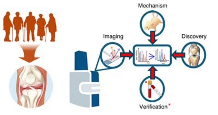
Bone Molecular Maps to Transform Early Osteoarthritis Detection
Osteoarthritis affects more than 500 million people worldwide and is a major cause of pain, disability, and reduced quality of life. By the time it is diagnosed through symptoms and visible cartilage loss,... Read moreHematology
view channel
New Guidelines Aim to Improve AL Amyloidosis Diagnosis
Light chain (AL) amyloidosis is a rare, life-threatening bone marrow disorder in which abnormal amyloid proteins accumulate in organs. Approximately 3,260 people in the United States are diagnosed... Read more
Fast and Easy Test Could Revolutionize Blood Transfusions
Blood transfusions are a cornerstone of modern medicine, yet red blood cells can deteriorate quietly while sitting in cold storage for weeks. Although blood units have a fixed expiration date, cells from... Read more
Automated Hemostasis System Helps Labs of All Sizes Optimize Workflow
High-volume hemostasis sections must sustain rapid turnaround while managing reruns and reflex testing. Manual tube handling and preanalytical checks can strain staff time and increase opportunities for error.... Read more
High-Sensitivity Blood Test Improves Assessment of Clotting Risk in Heart Disease Patients
Blood clotting is essential for preventing bleeding, but even small imbalances can lead to serious conditions such as thrombosis or dangerous hemorrhage. In cardiovascular disease, clinicians often struggle... Read moreMicrobiology
view channel
CRISPR-Based Technology Neutralizes Antibiotic-Resistant Bacteria
Antibiotic resistance has accelerated into a global health crisis, with projections estimating more than 10 million deaths per year by 2050 as drug-resistant “superbugs” continue to spread.... Read more
Comprehensive Review Identifies Gut Microbiome Signatures Associated With Alzheimer’s Disease
Alzheimer’s disease affects approximately 6.7 million people in the United States and nearly 50 million worldwide, yet early cognitive decline remains difficult to characterize. Increasing evidence suggests... Read morePathology
view channel
AI-Powered Microscope Diagnoses Malaria in Blood Smears Within Minutes
Malaria remains one of the world’s deadliest infectious diseases, killing hundreds of thousands each year, mostly in under-resourced regions where laboratory infrastructure is limited. Diagnosis still... Read more
Engineered Yeast Cells Enable Rapid Testing of Cancer Immunotherapy
Developing new cancer immunotherapies is a slow, costly, and high-risk process, particularly for CAR T cell treatments that must precisely recognize cancer-specific antigens. Small differences in tumor... Read moreTechnology
view channel
Robotic Technology Unveiled for Automated Diagnostic Blood Draws
Routine diagnostic blood collection is a high‑volume task that can strain staffing and introduce human‑dependent variability, with downstream implications for sample quality and patient experience.... Read more
ADLM Launches First-of-Its-Kind Data Science Program for Laboratory Medicine Professionals
Clinical laboratories generate billions of test results each year, creating a treasure trove of data with the potential to support more personalized testing, improve operational efficiency, and enhance patient care.... Read moreAptamer Biosensor Technology to Transform Virus Detection
Rapid and reliable virus detection is essential for controlling outbreaks, from seasonal influenza to global pandemics such as COVID-19. Conventional diagnostic methods, including cell culture, antigen... Read more
AI Models Could Predict Pre-Eclampsia and Anemia Earlier Using Routine Blood Tests
Pre-eclampsia and anemia are major contributors to maternal and child mortality worldwide, together accounting for more than half a million deaths each year and leaving millions with long-term health complications.... Read moreIndustry
view channel
WHX Labs in Dubai spotlights leadership skills shaping next-generation laboratories
WHX Labs in Dubai (formerly Medlab Middle East), held at Dubai World Trade Centre (DWTC) from 10–13 February, brings together international experts to discuss the factors redefining laboratory leadership,... Read moreNew Collaboration Brings Automated Mass Spectrometry to Routine Laboratory Testing
Mass spectrometry is a powerful analytical technique that identifies and quantifies molecules based on their mass and electrical charge. Its high selectivity, sensitivity, and accuracy make it indispensable... Read more
AI-Powered Cervical Cancer Test Set for Major Rollout in Latin America
Noul Co., a Korean company specializing in AI-based blood and cancer diagnostics, announced it will supply its intelligence (AI)-based miLab CER cervical cancer diagnostic solution to Mexico under a multi‑year... Read more


