MALDI-TOF MS Identifies Oomycete Causing Pythiosis
|
By LabMedica International staff writers Posted on 12 Dec 2018 |

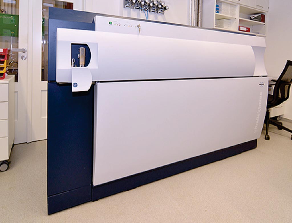
Image: The UltrafleXtreme MALDI-TOF/TOF mass spectrometer (Photo courtesy of Bruker Daltonics).
Pythiosis is an invasive, difficult-to-treat, life-threatening infectious disease caused by Pythium insidiosum, a member of the unique group of fungus-like microorganisms called oomycetes. The disease has been increasingly reported worldwide.
In the past decade, the matrix-assisted laser desorption ionization–time of flight mass spectrometry (MALDI-TOF MS) has emerged as a novel and powerful diagnostic tool for facilitating the clinical identification of many pathogenic microorganisms, including bacteria and fungi.
Scientists at the Mahidol University (Bangkok, Thailand) isolated a total of 13 strains of P. insidiosum, isolated from eight humans and five animals with pythiosis, from different geographic locations. All organisms were maintained on Sabouraud dextrose agar at 25 °C. Several small portions of a colony of each organism were transferred to a 50-mL flask containing 10 mL Sabouraud dextrose broth, and incubated at 37 °C for one week, before harvesting fungal material for protein extraction.
Protein was extracted from harvested organisms and was spotted onto a clean ground steel target plate in 40 replicates (for generating a MALDI-TOF MS database of P. insidiosum) or five replicates (for assessing the MALDI-TOF MS for identification of P. insidiosum), air dried at room temperature before being processed. After the matrix solution was air dried at room temperature, the sample was promptly analyzed, using a Bruker ultrafleXtreme mass spectrometer. Genomic DNA (gDNA) templates were extracted from the organisms and subjected to single nucleotide polymorphism-based multiplex polymerase chain reaction (PCR).
The team reported that the MALDI-TOF MS accurately identified all 13 P. insidiosum strains tested, at the species level. Mass spectra of P. insidiosum did not match any other microorganisms, including fungi (i.e., Aspergillus species, Fusarium species, and fungal species of the class Zygomycetes), which have similar microscopic morphologies with this oomycete. MALDI-TOF MS- and rDNA sequence-based biotyping methods consistently classified P. insidiosum into three groups: Clade-I (American strains), II (Asian and Australian strains), and III (mostly Thai strains).
The authors concluded that MALDI-TOF MS has been successfully used for identification and biotyping of P. insidiosum. The obtained mass spectral database allows clinical microbiology laboratories, well equipped with a MALDI-TOF mass spectrometer, to conveniently identify P. insidiosum, without requiring any pathogen-specific reagents (i.e., antigen, antibody or primers). The study was published in the December 2018 issue of the International Journal of Infectious Diseases.
Related Links:
Mahidol University
Latest Molecular Diagnostics News
- New Genome Sequencing Technique Measures Epstein-Barr Virus in Blood
- Blood Test Boosts Early Detection of Brain Cancer
- Molecular Monitoring Approach Helps Bladder Cancer Patients Avoid Surgery
- Genetic Tests to Speed Diagnosis of Lymphatic Disorders
- Changes In Lymphatic Vessels Can Aid Early Identification of Aggressive Oral Cancer
- New Extraction Kit Enables Consistent, Scalable cfDNA Isolation from Multiple Biofluids
- New CSF Liquid Biopsy Assay Reveals Genomic Insights for CNS Tumors
- AI-Powered Liquid Biopsy Classifies Pediatric Brain Tumors with High Accuracy
- Group A Strep Molecular Test Delivers Definitive Results at POC in 15 Minutes
- Rapid Molecular Test Identifies Sepsis Patients Most Likely to Have Positive Blood Cultures
- Light-Based Sensor Detects Early Molecular Signs of Cancer in Blood
- New Testing Method Predicts Trauma Patient Recovery Days in Advance
- Simple Method Predicts Risk of Brain Tumor Recurrence
- Genetic Test Could Improve Early Detection of Prostate Cancer
- Bone Molecular Maps to Transform Early Osteoarthritis Detection
- POC Testing for Hepatitis B DNA as Effective as Traditional Laboratory Testing
Channels
Clinical Chemistry
view channel
Simple Blood Test Offers New Path to Alzheimer’s Assessment in Primary Care
Timely evaluation of cognitive symptoms in primary care is often limited by restricted access to specialized diagnostics and invasive confirmatory procedures. Clinicians need accessible tools to determine... Read more
Existing Hospital Analyzers Can Identify Fake Liquid Medical Products
Counterfeit and substandard medicines remain a serious global health threat, with World Health Organization estimates suggesting that 10.5% of medicines in low- and middle-income countries are either fake... Read moreMolecular Diagnostics
view channel
New Genome Sequencing Technique Measures Epstein-Barr Virus in Blood
The Epstein–Barr virus (EBV) infects up to 95% of adults worldwide and remains in the body for life. While usually kept under control, the virus is linked to cancers such as Hodgkin’s lymphoma and autoimmune... Read more
Blood Test Boosts Early Detection of Brain Cancer
Brain and central nervous system (CNS) tumors are often diagnosed at an advanced stage, when treatment options are limited, and survival rates remain low. Around 300,000 new cases are diagnosed each year... Read moreHematology
view channel
Rapid Cartridge-Based Test Aims to Expand Access to Hemoglobin Disorder Diagnosis
Sickle cell disease and beta thalassemia are hemoglobin disorders that often require referral to specialized laboratories for definitive diagnosis, delaying results for patients and clinicians.... Read more
New Guidelines Aim to Improve AL Amyloidosis Diagnosis
Light chain (AL) amyloidosis is a rare, life-threatening bone marrow disorder in which abnormal amyloid proteins accumulate in organs. Approximately 3,260 people in the United States are diagnosed... Read moreImmunology
view channel
New Biomarker Predicts Chemotherapy Response in Triple-Negative Breast Cancer
Triple-negative breast cancer is an aggressive form of breast cancer in which patients often show widely varying responses to chemotherapy. Predicting who will benefit from treatment remains challenging,... Read moreBlood Test Identifies Lung Cancer Patients Who Can Benefit from Immunotherapy Drug
Small cell lung cancer (SCLC) is an aggressive disease with limited treatment options, and even newly approved immunotherapies do not benefit all patients. While immunotherapy can extend survival for some,... Read more
Whole-Genome Sequencing Approach Identifies Cancer Patients Benefitting From PARP-Inhibitor Treatment
Targeted cancer therapies such as PARP inhibitors can be highly effective, but only for patients whose tumors carry specific DNA repair defects. Identifying these patients accurately remains challenging,... Read more
Ultrasensitive Liquid Biopsy Demonstrates Efficacy in Predicting Immunotherapy Response
Immunotherapy has transformed cancer treatment, but only a small proportion of patients experience lasting benefit, with response rates often remaining between 10% and 20%. Clinicians currently lack reliable... Read morePathology
view channel
Single Sample Classifier Predicts Cancer-Associated Fibroblast Subtypes in Patient Samples
Pancreatic ductal adenocarcinoma (PDAC) remains one of the deadliest cancers, in part because of its dense tumor microenvironment that influences how tumors grow and respond to treatment.... Read more
New AI-Driven Platform Standardizes Tuberculosis Smear Microscopy Workflow
Sputum smear microscopy remains central to tuberculosis treatment monitoring and follow-up, particularly in high‑burden settings where serial testing is routine. Yet consistent, repeatable bacillary assessment... Read more
AI Tool Uses Blood Biomarkers to Predict Transplant Complications Before Symptoms Appear
Stem cell and bone marrow transplants can be lifesaving, but serious complications may arise months after patients leave the hospital. One of the most dangerous is chronic graft-versus-host disease, in... Read moreTechnology
view channel
Blood Test “Clocks” Predict Start of Alzheimer’s Symptoms
More than 7 million Americans live with Alzheimer’s disease, and related health and long-term care costs are projected to reach nearly USD 400 billion in 2025. The disease has no cure, and symptoms often... Read more
AI-Powered Biomarker Predicts Liver Cancer Risk
Liver cancer, or hepatocellular carcinoma, causes more than 800,000 deaths worldwide each year and often goes undetected until late stages. Even after treatment, recurrence rates reach 70% to 80%, contributing... Read more
Robotic Technology Unveiled for Automated Diagnostic Blood Draws
Routine diagnostic blood collection is a high‑volume task that can strain staffing and introduce human‑dependent variability, with downstream implications for sample quality and patient experience.... Read more
ADLM Launches First-of-Its-Kind Data Science Program for Laboratory Medicine Professionals
Clinical laboratories generate billions of test results each year, creating a treasure trove of data with the potential to support more personalized testing, improve operational efficiency, and enhance patient care.... Read moreIndustry
view channel
QuidelOrtho Collaborates with Lifotronic to Expand Global Immunoassay Portfolio
QuidelOrtho (San Diego, CA, USA) has entered a long-term strategic supply agreement with Lifotronic Technology (Shenzhen, China) to expand its global immunoassay portfolio and accelerate customer access... Read more


