Simple Test Predicts Childhood Cancer Relapse
|
By LabMedica International staff writers Posted on 26 May 2016 |


|
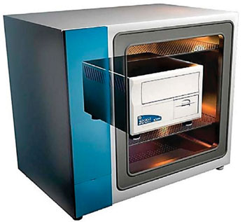
Image: The IncuCyte zoom continuous live-cell imaging and analysis system (Photo courtesy of Essen Biosciences).
High-risk neuroblastoma, which occurs mostly in children under five, is treated with surgery and chemotherapy, and neuroblastomas currently rated low risk are just removed or left untreated while doctors 'wait and see', but a fraction of low risk tumors recur and ultimately kill.
A cheap simple test could accurately predict the recurrence of a childhood cancer, as a protein marker has been pinpointed which when absent, shows neuroblastoma is almost certain to recur. It means children with low-risk neuroblastoma, who do not have the biomarker, can be reclassified as at high risk of relapse and have chemotherapy earlier.
Scientists at Brunel University London (UK) and their colleagues collected a retrospective series of primary tumors from neuroblastoma (NB) patients. Formalin-fixed, paraffin-embedded tissue sections from NB tumors were studied. Each tumor area tested contained malignant cells, assessed by histological examination. Quantification of immunofluorescence- or DAB-positive tumor cells was performed on serial tumor tissue sections. Tumor cells were distinguished in the samples using NB-specific marker, the homophilic binding glycoprotein neural cell adhesion molecule (NCAM, CD56).
The scientists carried out cell proliferation assays using IncuCyte live-cell imaging system (Essen Biosciences, Ann Arbor, MI, USA); immunostaining of cultured cells; Western blotting; angiogenesis assays where the images were captured using JuLI smart fluorescent cell analyzer (Baker Ruskinn, Sanford, ME; USA). The investigators used several other methodologies to identify the Promyelocytic Leukaemia protein-1(PML-1) and gene expression was assessed using chip microarrays.
PML was detected in the developing and adult sympathetic nervous system, whereas it was not expressed or low in metastatic neuroblastoma tumors. Reduced PML expression in patients with low-risk cancers, i.e. localized and negative for the V-Myc Avian Myelocytomatosis Viral Oncogene Neuroblastoma Derived Homolog (MYCN) protooncogene was strongly associated with tumor recurrence. PML-I, but not PML-IV, isoform suppresses angiogenesis via upregulation of thrombospondin-2 (TSP-2), a key inhibitor of angiogenesis. Finally, PML-I and TSP-2 expression inversely correlates with tumor angiogenesis and recurrence in localized neuroblastomas.
Paolo Salomoni, PhD, a professor and senior author of the study said, “We have found the absence of PML a very precise marker of tumor recurrence. In the low risk tumors, the absence of PML will be a very useful marker. What we can now say is that even some tumors classified as low risk, that would have previously gone untreated, if they show no expression of PML, they ought now to be classified as high risk.” The study was published on April 13, 2016, in the journal Clinical Cancer Research.
Related Links:
Brunel University London
Essen Biosciences
Baker Ruskinn





A cheap simple test could accurately predict the recurrence of a childhood cancer, as a protein marker has been pinpointed which when absent, shows neuroblastoma is almost certain to recur. It means children with low-risk neuroblastoma, who do not have the biomarker, can be reclassified as at high risk of relapse and have chemotherapy earlier.
Scientists at Brunel University London (UK) and their colleagues collected a retrospective series of primary tumors from neuroblastoma (NB) patients. Formalin-fixed, paraffin-embedded tissue sections from NB tumors were studied. Each tumor area tested contained malignant cells, assessed by histological examination. Quantification of immunofluorescence- or DAB-positive tumor cells was performed on serial tumor tissue sections. Tumor cells were distinguished in the samples using NB-specific marker, the homophilic binding glycoprotein neural cell adhesion molecule (NCAM, CD56).
The scientists carried out cell proliferation assays using IncuCyte live-cell imaging system (Essen Biosciences, Ann Arbor, MI, USA); immunostaining of cultured cells; Western blotting; angiogenesis assays where the images were captured using JuLI smart fluorescent cell analyzer (Baker Ruskinn, Sanford, ME; USA). The investigators used several other methodologies to identify the Promyelocytic Leukaemia protein-1(PML-1) and gene expression was assessed using chip microarrays.
PML was detected in the developing and adult sympathetic nervous system, whereas it was not expressed or low in metastatic neuroblastoma tumors. Reduced PML expression in patients with low-risk cancers, i.e. localized and negative for the V-Myc Avian Myelocytomatosis Viral Oncogene Neuroblastoma Derived Homolog (MYCN) protooncogene was strongly associated with tumor recurrence. PML-I, but not PML-IV, isoform suppresses angiogenesis via upregulation of thrombospondin-2 (TSP-2), a key inhibitor of angiogenesis. Finally, PML-I and TSP-2 expression inversely correlates with tumor angiogenesis and recurrence in localized neuroblastomas.
Paolo Salomoni, PhD, a professor and senior author of the study said, “We have found the absence of PML a very precise marker of tumor recurrence. In the low risk tumors, the absence of PML will be a very useful marker. What we can now say is that even some tumors classified as low risk, that would have previously gone untreated, if they show no expression of PML, they ought now to be classified as high risk.” The study was published on April 13, 2016, in the journal Clinical Cancer Research.
Related Links:
Brunel University London
Essen Biosciences
Baker Ruskinn

Platinum Member
Automated Coagulation Analyzer
Hemolumi H6
Manual Pipetting Aid
Pipette Controllers macro
Gold Member
Quantitative POC Immunoassay Analyzer
EASY READER+
Latest Molecular Diagnostics News
- Streamlined Genetic Testing Speeds Pediatric Immune Disorder Diagnosis
- Clinical Trial Evaluates Liquid Biopsy for Early-Stage Breast Cancer Detection
- RNA-Based Test Predicts Immunotherapy Response in Non-Small Cell Lung Cancer
- Modeling Study Shows Blood-Based CRC Screening Could Prevent More Deaths Than Stool Tests
- Noninvasive Cervical Test Detects Endometrial Cancer for Triage
- AI Tool Classifies Pathogenic Variants in Severe Childhood Epilepsy
- Molecular Test Detects Barrett’s Esophagus to Guide Endoscopy Referral
- Blood-Based Metagenomic Test Detects Wide Range of Pathogens from Single Sample
- New RNA Detection Tool Shows Promise for HIV and Other Viral Testing
- Tissue-Specific Vesicle Isolation Expands Molecular Insights Into Liver Disease
- Whole Genome Sequencing Solution Targets Unsolved Rare Disease Cases
- Validated Test Predicts Recurrence Risk and Radiation Benefit in Early-Stage Breast Cancer
- Study Shows How Genome Reference Bias Can Mislead Preventive Testing
- Genomic Screening Expands Detection of Treatable Conditions in Newborns
- Age-Based Genetic Testing May Miss Most Inherited Cancer Risk Variants
- Pooled Sputum Testing Offers Lower-Cost Molecular Diagnosis for Tuberculosis
Channels
Clinical Chemistry
view channel
Study Links Saliva-Based Epigenetic Signals to OCD Treatment Outcomes
Obsessive-compulsive disorder (OCD) causes unwanted, recurring thoughts and repetitive behaviors that can severely disrupt daily life. Exposure-based psychotherapy is among the most effective treatments,... Read more
New One-Step Syphilis Test Could Enable Earlier Detection and Faster Treatment
Syphilis can be difficult to diagnose because its symptoms may resemble other diseases and appear and disappear without treatment. Undetected infection can continue causing harm, spread to partners and... Read more

Molecular Diagnostics
view channel
Streamlined Genetic Testing Speeds Pediatric Immune Disorder Diagnosis
Children with genetic immune conditions can develop life-threatening illness, unusual infections, or autoimmune problems before the underlying cause is identified. More than 500 of these rare disorders... Read more
Clinical Trial Evaluates Liquid Biopsy for Early-Stage Breast Cancer Detection
Breast cancer screening relies primarily on mammography, but abnormal findings can lead to additional imaging, biopsy, or repeat imaging after six months, creating anxiety and potentially delaying treatment... Read more
RNA-Based Test Predicts Immunotherapy Response in Non-Small Cell Lung Cancer
Non-small cell lung cancer (NSCLC) represents the largest U.S. patient population eligible for immune checkpoint inhibitor therapy. Treatment selection often relies on PD-L1 expression, but a single protein... Read more
Modeling Study Shows Blood-Based CRC Screening Could Prevent More Deaths Than Stool Tests
Colorectal cancer (CRC) screening depends on patient participation and completion of follow-up colonoscopy after an abnormal noninvasive result. Access barriers can make both steps difficult, particularly... Read moreHematology
view channel
New Myelofibrosis Guidelines Recommend Expanded Genetic Testing at Diagnosis
Myelofibrosis is a rare, life-threatening blood cancer affecting up to six per 100,000 people. The disease causes scarring in the bone marrow, limiting the production of healthy blood cells and potentially... Read more
Handheld Point-of-Care Test Aims to Enable Rapid Coagulation Assessment in Trauma
Severe blood loss and inadequate tissue oxygenation are leading causes of preventable death after traumatic injury. Blood products are essential in trauma care, but cold-chain storage, short shelf life,... Read more

Immunology
view channel
Immune Profiling Initiative Aims to Improve Bone Marrow Analysis in Leukemia
Acute myeloid leukemia (AML) is an aggressive blood cancer in which assessing disease burden after therapy can help guide clinical evaluation. Patients with measurable residual disease (MRD) after treatment... Read more
Immune Profiling Identifies Early Maternal Autoantibodies Linked to Preterm Birth Risk
A typical pregnancy lasts about 40 weeks, but labor begins prematurely in roughly one in 10 pregnancies. Preterm birth is the leading cause of newborn death worldwide, yet its underlying causes and which... Read more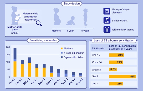
Longitudinal Blood Testing Reveals Changes in Allergen Sensitization During Childhood
Food allergy is a major childhood health concern, and distinguishing persistent risk from transient sensitization can guide clinical decisions. Specific IgE antibodies may be detectable even when a child... Read more
Microbiology
view channel
Molecular Panel Gains WHO Emergency Use Listing to Support Ebola Outbreak Response
The ongoing Ebola Bundibugyo outbreak in the Democratic Republic of the Congo and neighboring countries has become one of the largest outbreaks of this Ebola virus species recorded to date.... Read more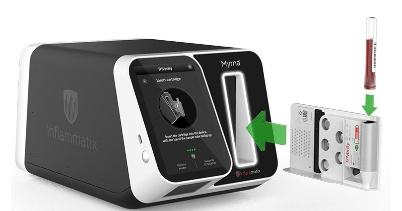
Rapid Host-Response Test Aims to Predict Corticosteroid Benefit in Sepsis
Sepsis and severe infections are difficult to treat because patients with similar clinical presentations may have very different underlying immune responses. Corticosteroids are commonly used in severe... Read more

Technology
view channel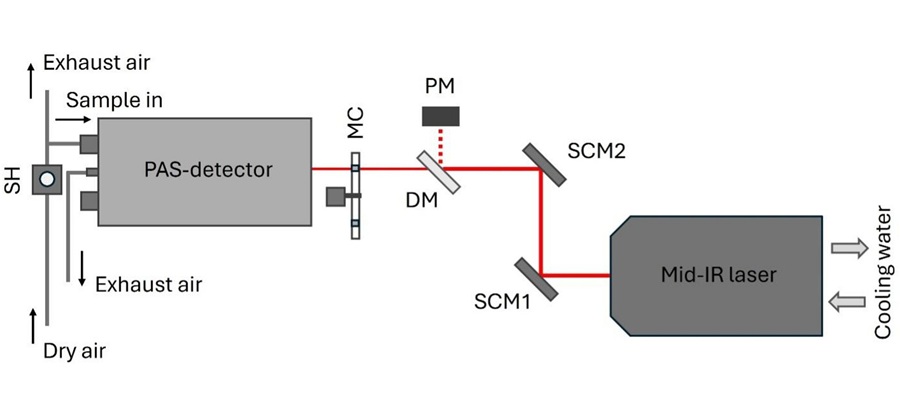
Laser-Based Swab Analysis Shows Promise for Detecting Disease-Linked Odor Patterns
Disease-related changes in volatile organic compounds can alter body odor, producing measurable patterns in exhaled breath and bodily fluids. Current analytical methods can be complex, time-consuming,... Read more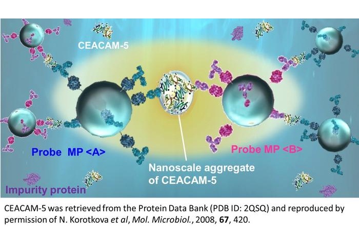
Laser-Enhanced Assay Boosts Sensitivity for Colorectal Cancer Biomarker Detection
Colorectal cancer is the third most commonly diagnosed cancer and the second leading cause of cancer-related death worldwide. Early detection remains critical, but cancer biomarkers can produce only faint... Read more

Industry
view channel
Hologic to Broaden Breast Cancer Testing Portfolio Through Reveal Genomics Acquisition
Hologic (Marlborough, MA, USA) has signed a definitive agreement to acquire Reveal Genomics (Barcelona, Spain), a precision oncology company developing genomic diagnostic tests to help personalize cancer treatment.... Read more