Blood Test Detects Chronic Traumatic Encephalopathy During Life
|
By LabMedica International staff writers Posted on 22 Mar 2016 |


|
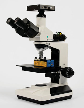
Image: The NanoSight LM10 instrument provides an easy-to-use, reproducible platform for nanoparticle characterization (Photo courtesy of Malvern Instruments).
Chronic traumatic encephalopathy (CTE) is a tauopathy associated with prior exposure to repetitive head impacts, such as those incurred through American football and other collision sports and diagnosis is made through neuropathological examination.
A method has been developed for measuring plasma exosomal tau. Exosomes are very small vesicles that are released from all types of cells throughout the body, including brain cells. They can be isolated in all body fluids, including plasma, a component of blood.
Scientists at Boston University School of Medicine (MA, USA) and their colleagues examined 78 former National Football League (NFL) players and a control group of 17 former non-contact sport athletes. Extracellular vesicles were isolated from plasma. Fluorescent nanoparticle tracking analysis was used to determine the number of vesicles staining positive for tau. Blood was drawn by venipuncture and immediately processed. Plasma samples were stored in 0.75 mL tubes at -80 °C, packed in dry ice, and shipped overnight in one batch to the Exosome Sciences laboratory (Monmouth Junction, NJ, USA).
Extracellular vesicles were isolated from the plasma samples by size exclusion chromatography. Plasma samples were applied to the column and it was then run isocratically. The elution was monitored at 280 nm and the void volume material (exosome fraction) was collected and pooled. The size profiles and concentrations of the chromatographically isolated exosomes were performed using the Nanosight LM10 instrument (Malvern Instruments Ltd.; Malvern, UK) equipped with a 405 nm laser, a scientific complementary metal-oxide-semiconductor (sCMOS) camera and Nanoparticle Tracking Analysis (NTA) software.
The method of measuring plasma exosomal tau is referred to as a TauSome. The groups did not differ in total plasma exosomes, but the NFL group had significantly higher plasma exosomal tau than the control group. The C-statistic is maximized when the plasma exosomal tau level is set to 0. Using 0 as a threshold resulted in 82% sensitivity, 100% specificity, 100% positive predictive value and 53% negative predictive value. The number of tau-positive plasma exosomes was significantly correlated with performance on standardized tests of memory and psychomotor speed; the higher the TauSome level, the worse the performance.
Jim Joyce, Founder of Exosome Sciences and Chairman and CEO of Aethlon Medical (San Diego, CA, USA), said, “We are extremely pleased that our initial study data has been published and we appreciate forthcoming opportunities to further advance our TauSome biomarker as a noninvasive solution to detect and monitor CTE in living individuals.” The study was published on February 10, 2016, in the Journal of Alzheimer's Disease.
Related Links:
Boston University School of Medicine
Exosome Sciences
Malvern Instruments Ltd.






A method has been developed for measuring plasma exosomal tau. Exosomes are very small vesicles that are released from all types of cells throughout the body, including brain cells. They can be isolated in all body fluids, including plasma, a component of blood.
Scientists at Boston University School of Medicine (MA, USA) and their colleagues examined 78 former National Football League (NFL) players and a control group of 17 former non-contact sport athletes. Extracellular vesicles were isolated from plasma. Fluorescent nanoparticle tracking analysis was used to determine the number of vesicles staining positive for tau. Blood was drawn by venipuncture and immediately processed. Plasma samples were stored in 0.75 mL tubes at -80 °C, packed in dry ice, and shipped overnight in one batch to the Exosome Sciences laboratory (Monmouth Junction, NJ, USA).
Extracellular vesicles were isolated from the plasma samples by size exclusion chromatography. Plasma samples were applied to the column and it was then run isocratically. The elution was monitored at 280 nm and the void volume material (exosome fraction) was collected and pooled. The size profiles and concentrations of the chromatographically isolated exosomes were performed using the Nanosight LM10 instrument (Malvern Instruments Ltd.; Malvern, UK) equipped with a 405 nm laser, a scientific complementary metal-oxide-semiconductor (sCMOS) camera and Nanoparticle Tracking Analysis (NTA) software.
The method of measuring plasma exosomal tau is referred to as a TauSome. The groups did not differ in total plasma exosomes, but the NFL group had significantly higher plasma exosomal tau than the control group. The C-statistic is maximized when the plasma exosomal tau level is set to 0. Using 0 as a threshold resulted in 82% sensitivity, 100% specificity, 100% positive predictive value and 53% negative predictive value. The number of tau-positive plasma exosomes was significantly correlated with performance on standardized tests of memory and psychomotor speed; the higher the TauSome level, the worse the performance.
Jim Joyce, Founder of Exosome Sciences and Chairman and CEO of Aethlon Medical (San Diego, CA, USA), said, “We are extremely pleased that our initial study data has been published and we appreciate forthcoming opportunities to further advance our TauSome biomarker as a noninvasive solution to detect and monitor CTE in living individuals.” The study was published on February 10, 2016, in the Journal of Alzheimer's Disease.
Related Links:
Boston University School of Medicine
Exosome Sciences
Malvern Instruments Ltd.

Platinum Member
Automated Coagulation Analyzer
Hemolumi H6
Portable POCT Blood Gas Analyzer
BD100
Gold Member
Platelet Function Analyzer
PL-12
Latest Clinical Chem. News
- Daily Blood Biomarker Testing May Help Guide Earlier Antibiotic Stopping in Sepsis
- Blood Proteins Reveal Signals Linked to Prostate Cancer Risk Years Before Diagnosis
- Two-Step Blood Testing Approach Reduces Uncertainty in Amyloid Assessment
- Multimodal Platform to Combine Blood and Imaging Biomarkers for Lung Nodule Assessment
- Study Links Saliva-Based Epigenetic Signals to OCD Treatment Outcomes
- New One-Step Syphilis Test Could Enable Earlier Detection and Faster Treatment
- Multimodal Clinical Data Models Predict Cachexia Risk Across Cancers
- Study Evaluates Alzheimer’s Blood Biomarker Across Racial and Ethnic Groups
- Blood Protein May Help Identify Treatment-Resistant Psychosis
- Electrochemical Biosensor Rapidly Detects Cerebrospinal Fluid Leaks
- Blood Protein Panel Supports Monitoring of Treated Celiac Disease
- Hemoglobin Levels May Help Signal Long-Term Dementia Risk
- Liquid Biopsy Platform Shows Promise for Early Gynecologic Cancer Detection
- Global Survey Finds Pediatric Troponin Testing Lacks Consistent Reference Intervals
- Multi-Omics Study Identifies Blood Marker for MASLD Progression
- Blood Biomarker Detects Alzheimer’s Changes Decades Before Symptoms in Down Syndrome
Channels
Clinical Chemistry
view channel
Daily Blood Biomarker Testing May Help Guide Earlier Antibiotic Stopping in Sepsis
Sepsis is a life-threatening condition caused by the body’s overwhelming response to infection and accounts for thousands of deaths each year. In intensive care, concern about stopping treatment too early... Read more
Blood Proteins Reveal Signals Linked to Prostate Cancer Risk Years Before Diagnosis
Prostate cancer is the most commonly diagnosed cancer in men worldwide. While many tumors grow slowly, aggressive forms can be life-threatening, underscoring the need for earlier risk identification.... Read more
Two-Step Blood Testing Approach Reduces Uncertainty in Amyloid Assessment
Blood-based biomarkers offer a more accessible, less expensive way to detect Alzheimer’s-related brain changes than traditional methods such as amyloid-beta positron emission tomography (Aβ-PET).... Read more
Multimodal Platform to Combine Blood and Imaging Biomarkers for Lung Nodule Assessment
Lung nodules are commonly detected during chest imaging and require careful risk assessment to determine whether follow-up imaging, additional testing, or biopsy is needed. With millions of patients evaluated... Read moreMolecular Diagnostics
view channel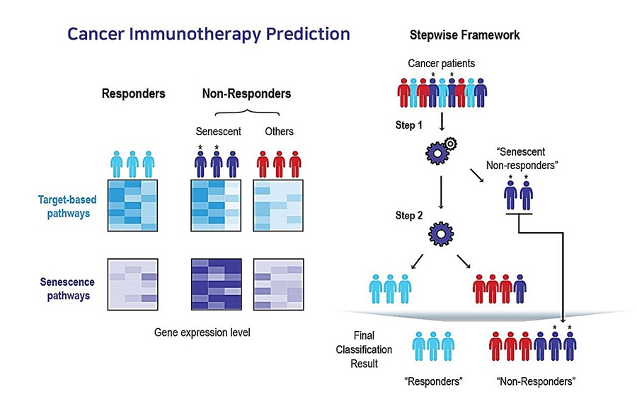
AI Tool Predicts Immunotherapy Response Using Tumor Senescence Signals
Patients receiving the same cancer immunotherapy can have markedly different outcomes, with some showing little or no response. Identifying these patients earlier could reduce the costs of ineffective... Read more
New Variant Tracking Option Expands Molecular Residual Disease Monitoring
Molecular residual disease (MRD) testing can detect traces of cancer that persist after treatment or emerge during recurrence. Because circulating tumor DNA (ctDNA) may be present at very low levels, residual... Read more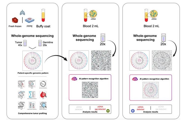

Hematology
view channel
New Myelofibrosis Guidelines Recommend Expanded Genetic Testing at Diagnosis
Myelofibrosis is a rare, life-threatening blood cancer affecting up to six per 100,000 people. The disease causes scarring in the bone marrow, limiting the production of healthy blood cells and potentially... Read more
Handheld Point-of-Care Test Aims to Enable Rapid Coagulation Assessment in Trauma
Severe blood loss and inadequate tissue oxygenation are leading causes of preventable death after traumatic injury. Blood products are essential in trauma care, but cold-chain storage, short shelf life,... Read more

Immunology
view channel
Antibody Profiling Predicts Inflammatory Bowel Disease Years Before Diagnosis
Inflammatory bowel disease (IBD), which includes Crohn’s disease and ulcerative colitis, involves abnormal immune responses that cause persistent inflammation and damage in the digestive system.... Read more
Immune Profiling Initiative Aims to Improve Bone Marrow Analysis in Leukemia
Acute myeloid leukemia (AML) is an aggressive blood cancer in which assessing disease burden after therapy can help guide clinical evaluation. Patients with measurable residual disease (MRD) after treatment... Read more
Immune Profiling Identifies Early Maternal Autoantibodies Linked to Preterm Birth Risk
A typical pregnancy lasts about 40 weeks, but labor begins prematurely in roughly one in 10 pregnancies. Preterm birth is the leading cause of newborn death worldwide, yet its underlying causes and which... Read more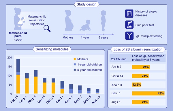
Longitudinal Blood Testing Reveals Changes in Allergen Sensitization During Childhood
Food allergy is a major childhood health concern, and distinguishing persistent risk from transient sensitization can guide clinical decisions. Specific IgE antibodies may be detectable even when a child... Read moreMicrobiology
view channel
Molecular Panel Gains WHO Emergency Use Listing to Support Ebola Outbreak Response
The ongoing Ebola Bundibugyo outbreak in the Democratic Republic of the Congo and neighboring countries has become one of the largest outbreaks of this Ebola virus species recorded to date.... Read more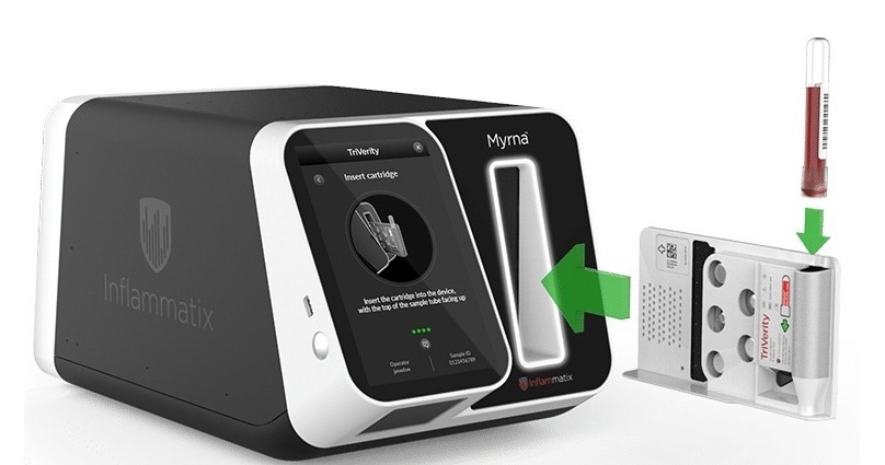
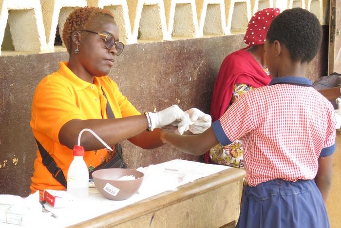
Rapid Host-Response Test Aims to Predict Corticosteroid Benefit in Sepsis
Sepsis and severe infections are difficult to treat because patients with similar clinical presentations may have very different underlying immune responses. Corticosteroids are commonly used in severe... Read more
Technology
view channel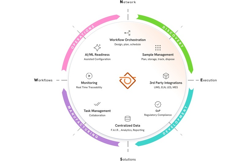
New AI-Powered System Aims to Streamline Laboratory and Manufacturing Workflows
Scientific laboratories supporting drug development, biotechnology, and materials research increasingly rely on coordinated digital workflows to move data, samples, and activities across teams.... Read more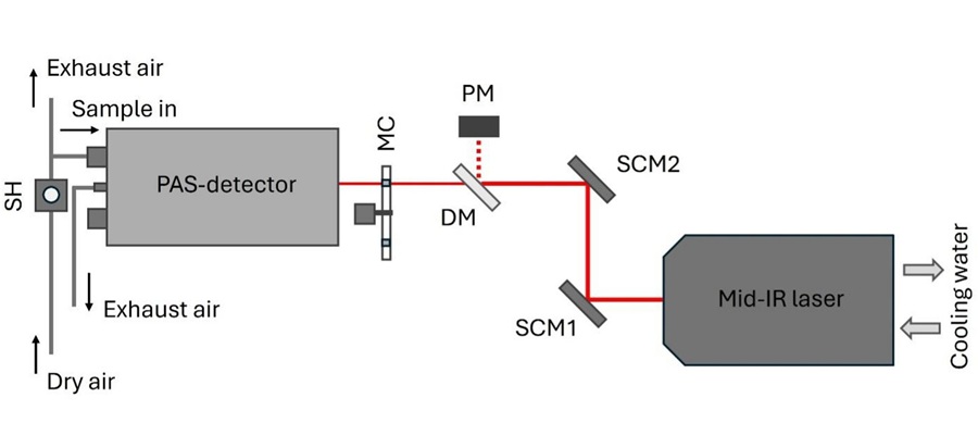
Laser-Based Swab Analysis Shows Promise for Detecting Disease-Linked Odor Patterns
Disease-related changes in volatile organic compounds can alter body odor, producing measurable patterns in exhaled breath and bodily fluids. Current analytical methods can be complex, time-consuming,... Read more
Industry
view channel
Collaboration to Expand Cervical Cancer Screening Access With At-Home HPV Collection
Cervical cancer is largely preventable with regular screening and early detection of human papillomavirus (HPV), the leading cause of the disease. Screening gaps persist, in part because traditional sample... Read more
Alzheimer’s Progression Blood Test Gains U.S. Patent Ahead of Planned Launch
Alzheimer’s disease is the most common cause of dementia, accounting for an estimated 60–70% of cases worldwide. Its progressive effects on memory, thinking, behavior, and daily functioning underscore... Read more