Lethal Prion Protein Causes Neuronal Death
|
By LabMedica International staff writers Posted on 01 Mar 2012 |


|
A single prion protein that is at least 10 times more lethal than larger prion species has been identified that causes disease similar to mad cow disease.
The toxic form of abnormal prion protein, known as TPrP, triggered various forms of neuronal damage, including molecular signatures very similar to that seen in the brains of prion-infected animal with bovine spongiform encephalopathy (BSE).
Scientists at the Scripps Research Institute (Jupiter, FL, USA) found that the most lethal form of prion protein was a specific structure known as alpha-helical. This toxic single molecule or "monomer" tests the existing theory that neuronal damage is associated with the toxicity of prion protein aggregates called "oligomers."
A variety of techniques was used to provide fresh insights into prion diseases, such as BSE and a rare human form Creutzfeldt-Jakob disease. Furthermore, it opened the possibility that associated neurotoxic proteins may play a role in neurodegenerative disorders, such as Parkinson, and Alzheimer's diseases.
In prion disease, proteinaceous infectious particles (infectious prions), believed to be made up only of protein, are able to reproduce, even though they lack ribonucleic acid (RNA) and DNA. Usually, mammalian cells generate cellular prion protein (PrP), when infected with a prion disease; the abnormal protein converts the normal host prion protein into its disease form.
Corinne Lasmézas, PhD, the lead author and a professor at Scripps, said "By identifying a single molecule as the most toxic species of prion proteins, we've opened a new chapter in understanding how prion-induced neurodegeneration occurs. We didn't think we would find neuronal death from this toxic monomer so close to what normally happens in the disease state. Now we have a powerful tool to explore the mechanisms of neurodegeneration."
Professor Lasmézas added, "Until now, it was thought that oligomers of proteins are toxic in all these disease. Since we found for the first time that an abnormally folded monomer is highly toxic, it opens up the possibility that this might be true also for some other protein misfolding diseases as well." The study was published on February 7, 2012 in the Proceedings of the National Academy of Sciences of the United States of America (PNAS).
Related Links:
Scripps Research Institute





The toxic form of abnormal prion protein, known as TPrP, triggered various forms of neuronal damage, including molecular signatures very similar to that seen in the brains of prion-infected animal with bovine spongiform encephalopathy (BSE).
Scientists at the Scripps Research Institute (Jupiter, FL, USA) found that the most lethal form of prion protein was a specific structure known as alpha-helical. This toxic single molecule or "monomer" tests the existing theory that neuronal damage is associated with the toxicity of prion protein aggregates called "oligomers."
A variety of techniques was used to provide fresh insights into prion diseases, such as BSE and a rare human form Creutzfeldt-Jakob disease. Furthermore, it opened the possibility that associated neurotoxic proteins may play a role in neurodegenerative disorders, such as Parkinson, and Alzheimer's diseases.
In prion disease, proteinaceous infectious particles (infectious prions), believed to be made up only of protein, are able to reproduce, even though they lack ribonucleic acid (RNA) and DNA. Usually, mammalian cells generate cellular prion protein (PrP), when infected with a prion disease; the abnormal protein converts the normal host prion protein into its disease form.
Corinne Lasmézas, PhD, the lead author and a professor at Scripps, said "By identifying a single molecule as the most toxic species of prion proteins, we've opened a new chapter in understanding how prion-induced neurodegeneration occurs. We didn't think we would find neuronal death from this toxic monomer so close to what normally happens in the disease state. Now we have a powerful tool to explore the mechanisms of neurodegeneration."
Professor Lasmézas added, "Until now, it was thought that oligomers of proteins are toxic in all these disease. Since we found for the first time that an abnormally folded monomer is highly toxic, it opens up the possibility that this might be true also for some other protein misfolding diseases as well." The study was published on February 7, 2012 in the Proceedings of the National Academy of Sciences of the United States of America (PNAS).
Related Links:
Scripps Research Institute

Platinum Member
Automated Coagulation Analyzer
Hemolumi H6
CMV CLIA Diagnostic
CLIA CMV IgA Screen Group
Gold Member
Nucleic Acid Extractor System
NEOS-96 XT
Latest Microbiology News
- Molecular Panel Gains WHO Emergency Use Listing to Support Ebola Outbreak Response
- Rapid Host-Response Test Aims to Predict Corticosteroid Benefit in Sepsis
- Oral and Lung Microbiome Changes Linked to Severe Long Covid Symptoms
- Study Finds Distinct Malaria Parasites Linked to Sickle Cell Trait Protection
- Rapid Urine Test Aids Diagnosis of Invasive Aspergillosis
- Nanodroplet CRISPR Technology Supports Rapid, Multiplexed Mycobacterial Identification
- Genomic Workflow Identifies Fungal Pathogens Before Blood Cultures Turn Positive
- FDA-Cleared Multiplex PCR Test Detects 13 Respiratory Pathogens in a Single Sample
- One-Hour Molecular Panel Expands Bloodstream Infection Testing for Gram-Negative Pathogens
- FDA Clears Rapid Phenotypic Antimicrobial Susceptibility System for Positive Blood Cultures
- New Urine Test Expands Mycotoxin Analysis to 31 Markers for Broader Exposure Assessment
- Collaboration Targets Blood-Based Ebola Detection Outside Central Laboratories
- Study Finds Frequent Overlap in Antibodies to Lyme and Other Tick-Borne Pathogens
- Early-Life Gut Microbiome Changes May Help Assess Type 1 Diabetes Risk
- FDA Breakthrough-Designated Urine Panel Aims to Shorten Pathogen and Susceptibility Testing
- Expanded Newborn Screening Program Increases Detection of Congenital Cytomegalovirus
Channels
Clinical Chemistry
view channel
Point-of-Care Blood Biomarker Test Detects Brain Injury Missed by CT
Traumatic brain injury remains difficult to assess in emergency settings because some patients continue to have symptoms despite normal computed tomography findings. Approximately 2.8 million people in the U.... Read more
Daily Blood Testing May Help Guide Earlier Antibiotic Stopping in Sepsis
Sepsis is a life-threatening condition caused by the body’s overwhelming response to infection and accounts for thousands of deaths each year. In intensive care, concern about stopping treatment too early... Read more

Molecular Diagnostics
view channel
Molecular Vaginitis Panel Gains FDA Clearance and CLIA Waiver for Point-of-Care Use
Vaginitis affects most women at least once in their lifetime and can cause painful, disruptive symptoms. Common causes include bacterial vaginosis, vulvovaginal candidiasis, and trichomoniasis, reflecting... Read more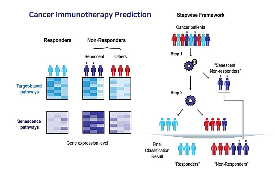
AI Tool Predicts Immunotherapy Response Using Tumor Senescence Signals
Patients receiving the same cancer immunotherapy can have markedly different outcomes, with some showing little or no response. Identifying these patients earlier could reduce the costs of ineffective... Read more
Hematology
view channel
New Myelofibrosis Guidelines Recommend Expanded Genetic Testing at Diagnosis
Myelofibrosis is a rare, life-threatening blood cancer affecting up to six per 100,000 people. The disease causes scarring in the bone marrow, limiting the production of healthy blood cells and potentially... Read more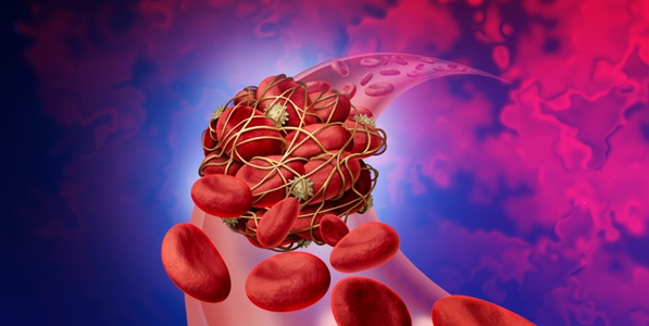
Handheld Point-of-Care Test Aims to Enable Rapid Coagulation Assessment in Trauma
Severe blood loss and inadequate tissue oxygenation are leading causes of preventable death after traumatic injury. Blood products are essential in trauma care, but cold-chain storage, short shelf life,... Read more

Immunology
view channel
Antibody Profiling Predicts Inflammatory Bowel Disease Years Before Diagnosis
Inflammatory bowel disease (IBD), which includes Crohn’s disease and ulcerative colitis, involves abnormal immune responses that cause persistent inflammation and damage in the digestive system.... Read more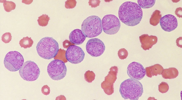
Immune Profiling Initiative Aims to Improve Bone Marrow Analysis in Leukemia
Acute myeloid leukemia (AML) is an aggressive blood cancer in which assessing disease burden after therapy can help guide clinical evaluation. Patients with measurable residual disease (MRD) after treatment... Read more
Immune Profiling Identifies Early Maternal Autoantibodies Linked to Preterm Birth Risk
A typical pregnancy lasts about 40 weeks, but labor begins prematurely in roughly one in 10 pregnancies. Preterm birth is the leading cause of newborn death worldwide, yet its underlying causes and which... Read more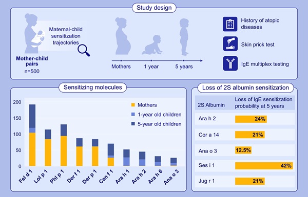
Longitudinal Blood Testing Reveals Changes in Allergen Sensitization During Childhood
Food allergy is a major childhood health concern, and distinguishing persistent risk from transient sensitization can guide clinical decisions. Specific IgE antibodies may be detectable even when a child... Read morePathology
view channel
Digital Pathology Test Stratifies Recurrence Risk in Early Breast Cancer
In hormone receptor-positive, human epidermal growth factor receptor 2 (HER2)-negative early breast cancer, accurate recurrence-risk assessment helps guide treatment decisions. However, individual risks... Read more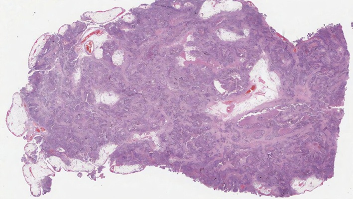
AI Pathology Tool Predicts DNA Repair Deficiency in High-Grade Serous Ovarian Cancer
High-grade serous tubo-ovarian carcinoma is an aggressive cancer arising in the ovaries or fallopian tubes. Some tumors exhibit homologous recombination deficiency (HRD), a feature reflecting impaired DNA repair.... Read more
Technology
view channel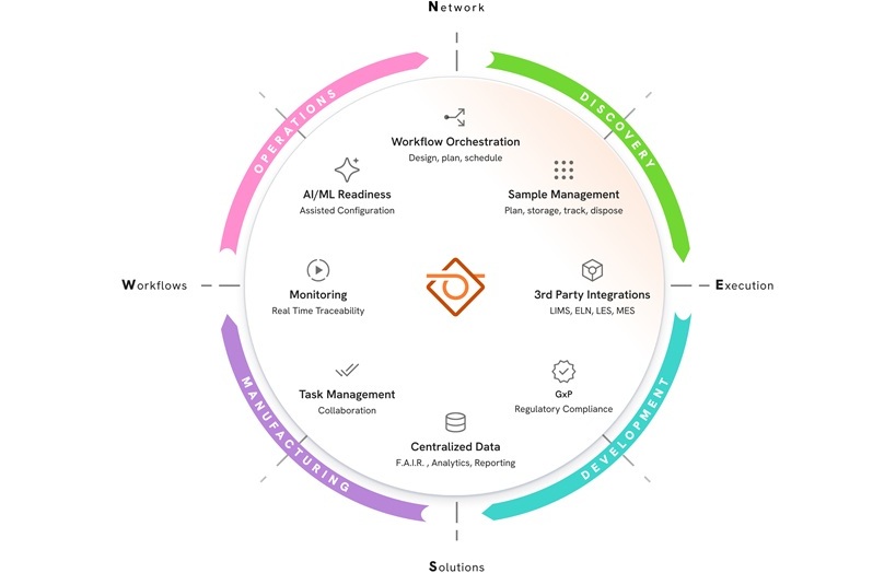
New AI-Powered System Aims to Streamline Laboratory and Manufacturing Workflows
Scientific laboratories supporting drug development, biotechnology, and materials research increasingly rely on coordinated digital workflows to move data, samples, and activities across teams.... Read more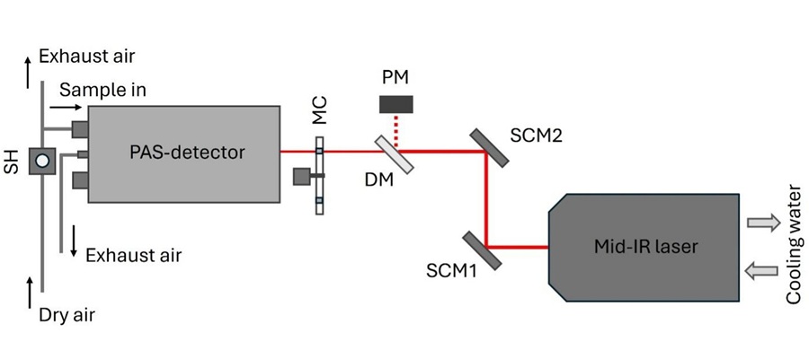
Laser-Based Swab Analysis Shows Promise for Detecting Disease-Linked Odor Patterns
Disease-related changes in volatile organic compounds can alter body odor, producing measurable patterns in exhaled breath and bodily fluids. Current analytical methods can be complex, time-consuming,... Read more
Industry
view channel
Qiagen Agrees to Go Private Through EQT-Led Acquisition
QIAGEN N.V. (Venlo, the Netherlands) and EQT Private Equity (Stockholm, Sweden) have agreed to an all-cash public takeover offer for all outstanding QIAGEN shares at EUR 46.50 per share.... Read more
Hologic and Inpeco Partner to Advance Molecular Laboratory Automation in Europe
Hologic, Inc. (Marlborough, MA, USA) and Inpeco S.A. (Novazzano, Switzerland) have signed a co-marketing partnership in Europe for Inpeco’s recently launched FlexLab X automation solution and Hologic’s... Read more