Paper-Based TB Test Could Increase Diagnoses Worldwide
By LabMedica International staff writers
Posted on 26 Sep 2017
In resource-limited areas, equipment requirements and long wait times for results are obstacles to early diagnosis and treatment of tuberculosis (TB). To tackle this problem, scientists have developed a rapid colorimetric TB assay using gold nanoparticles on a paper-based device than enables results to be read with a smartphone.Posted on 26 Sep 2017
Most of deaths from TB occur in low- and middle-income countries and remote areas. Conventional methods such as sputum smear microscopy, chest X-rays, and molecular-based tests require equipment, electricity, and specialized personnel. So lead researcher Chien-Fu Chen, professor at National Taiwan University (Taiwan), and colleagues from Chang Gung Memorial Hospital (Taiwan) and elsewhere in Taiwan, set out to come up with a more practical diagnostic test that can be read with a smartphone, a technology that is becoming increasingly available in emerging economies.
.")
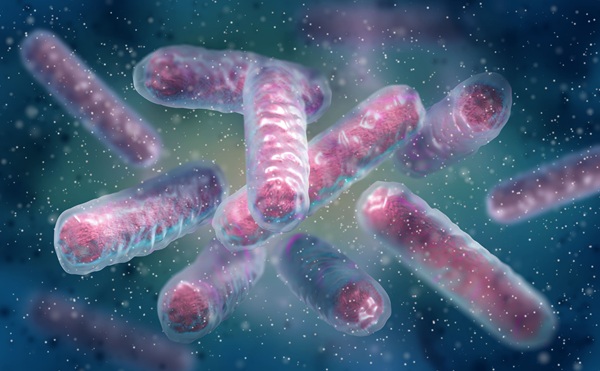
Image: Scientists have developed a rapid, paper-based TB text that allows results to be read with a smartphone (Photo courtesy of Wikimedia Commons).
The researchers combined gold nanoparticles with single-stranded DNA (ssDNA) sequences that bind to the genetic material of Mycobacterium tuberculosis. These nanoparticles were then incorporated into a paper-based device. Adding even a minute amount of lab-derived, double-stranded DNA (dsDNS) from M. tuberculosis changed the color of the test spots within an hour. Furthermore, multiple tests could be performed simultaneously with a 60 min turnaround time.
A smartphone camera was used to analyze the color change to determine the bacterial concentration. The researchers also tested a tissue sample from an infected patient to further demonstrate that the device could be used in the field.
The study, by Tsai TT et al, was published September 13, 2017, in the journal ACS Sensors.
Related Links:
National Taiwan University
Chang Gung Memorial Hospital

Gold Member
Neonatal Heel Incision Device
Tenderfoot


HIV-1 Molecular Diagnostic Assay
AltoStar HIV RT-PCR Kit 1.5

Urine Analyzer
respons® UDS100