Three Methods Evaluated for Malaria Detection
By LabMedica International staff writers
Posted on 23 May 2017
Malaria is a debilitating disease with high morbidity and mortality in Africa, commonly caused by different species of the genus Plasmodium in humans. Misdiagnosis is a major challenge in endemic areas because of other disease complications and technical expertise of the medical laboratory staff.Posted on 23 May 2017
The microscopic examination of Giemsa-stained thick and thin blood films has been used for the laboratory diagnosis of malaria for many years. The limitation of microscopy led to the development of reliable, easy-to-perform rapid diagnostic tests (RDTs) to detect the presence of malaria parasites at levels of accuracy compared to skilled microscopists.
.")
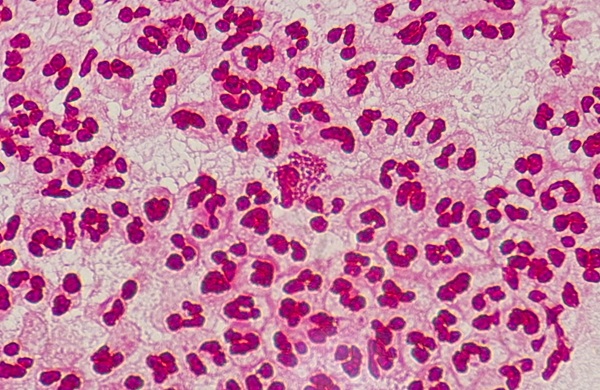
Image: Malaria parasites in a red blood cell (Photo courtesy of SPL).
Scientists at the Federal University Ndufu-Alike Ikwo and their colleagues used simple random sampling of a populations comprised of individuals who had shown clinical signs of malaria and in whom test for malaria parasite have been requested for by clinicians. The subject population included persons of different age groups such as children, adults and pregnant women. Peripheral blood samples were collected by finger prick and by venipuncture.
The study participants were screened for malaria parasites using Giemsa-stained malaria microscopy, three RDT kits, among the specimens collected, a total of 50 were randomly selected for nested polymerase chain reaction (PCR). Three different commercially available RDT kits for malaria parasites having different sensitivity and specificity were used to detect malaria parasites in the blood samples. These were; Carestart, SD Bioline PF and SD Bioline PF/PV. Molecular analysis was performed using a PCR technique based on amplification of 18s rRNA genes.
The team reported that malaria parasite was detected in 109/420 (25.95%) of the subjects by microscopy and all the species detected by microscopy were observed to be P. falciparum. Among the subjects studied, 96 (22.9%), 64 (15.2%) and 230 (54.8%) were positive by Carestart, SD Bioline PF and SD Bioline PF/PV respectively. Among the specimens that were subjected to molecular analysis, 16 (32.0%) were positive while 34 (68.0%) were negative for Plasmodium genes. Plasmodium falciparum was observed in all positive cases, P. malariae was present in 5/16 cases while P. ovale was present in 1/16 subject. Both of the latter species were present as co-infection with P. falciparum in all the subjects they were found.
The authors concluded that microscopy is still a good method for the diagnosis of malaria and having a good measure of agreement with PCR. Hence it is recommended that the laboratory diagnosis of malaria be performed using both microscopy and RDT of high sensitivity and specificity. The combination of these methods will ensure that laboratory reports on malaria diagnosis are of very high accuracy. The study was published on May 6, 2017, in the Malaria Journal.
.jpg)
Gold Member
Clinical Chemistry Assay
Sorbitol Dehydrogenase (SDH)


LAIR2 Antibody Pair Set
LAIR2 Antibody Pair [Biotin]

Pipette Calibration System
Artel PCS®