Two Classes of Fibroblasts Inhabit the Synovial Membrane in RA
By Gerald M. Slutzky, PhD
Posted on 07 Dec 2016
Rheumatoid arthritis researchers have identified two distinct classes of synovial fibroblasts (SFs) differentiated by expression of either PDPN (podoplanin) or CD248 (endosialin), which were located within different anatomical compartments of the inflamed synovial membrane.Posted on 07 Dec 2016
Investigators at the University of Birmingham (United Kingdom) studied two distinct SF populations that were located preferentially in the lining or sub-lining layers of the synovial membrane, which were defined by their expression of either PDPN or CD248, and explored their ability to undergo self-assembly and transmigration in vivo.
.")
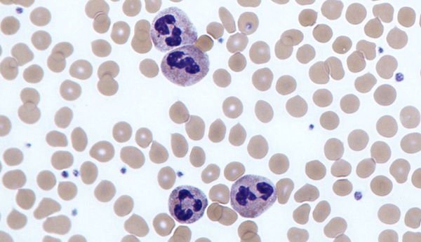
Image: A representation of the different cell types in the rheumatoid synovium (Photo courtesy of the University of Birmingham).
For this study SFs were cultured in vitro, and phenotypic changes following stimulation with interleukin (IL)-1beta, tumor necrosis factor (TNF)-alpha, and transforming growth factor (TGF)-beta1 were evaluated. To examine the phenotype of SF in vivo, a severe combined immunodeficiency (SCID) human-mouse model of cartilage destruction was utilized.
Results published in the November 18, 2016, online edition of the journal Arthritis Research and Therapy revealed that SFs in the lining layer in rheumatoid arthritis expressed high levels of PDPN compared to the normal synovium, whereas CD248 expression was restricted to sub-lining layer cells. TNF-alpha or IL1 stimulation in vitro resulted in an increased expression of PDPN. In contrast, stimulation with TGF-beta1 induced CD248 expression. The PDPN-expressing cells were associated with early fibroblast migration and cartilage erosion.
In the SCID human-mouse model, rheumatoid SF recapitulated the expression of PDPN and CD248. Thus, fibroblasts adjacent to cartilage expressed PDPN, and attached to, invaded, and degraded cartilage. Since PDPN-expressing cells were associated with early fibroblast migration and cartilage erosion in vivo, the investigators proposed that PDPN-expressing cells may be an attractive therapeutic target in rheumatoid arthritis.
First author Dr. Adam Croft, researcher in the rheumatology research group at the University of Birmingham, said, "This study not only shows the existence of distinct sub-sets of synovial fibroblasts, but also suggests that these cells are able to self-organize into lining and sub-lining layers in the presence of cartilage. Combined with the difference in migration rates between the two types of cell, these results are extremely promising in terms of finding new therapeutic targets for treatment of rheumatoid arthritis."
Related Links:
University of Birmingham

Gold Member
Automatic Hematology Analyzer
CF9600


Rapid Sepsis Test
SeptiCyte RAPID

New
Food Allergy Screening ELISA Kit
Allerquant 14G B ELISA