Novel 3D Printing Technique Installs NIV Sensors in Lab-on-a Chip Devices
By LabMedica International staff writers
Posted on 04 Nov 2016
A team of biomedical engineers developed an advanced three-dimensional (3D) printing technique to embed noninvasive strain sensors into living tissue as an integral component of organ-on-a-chip devices.Posted on 04 Nov 2016
Microphysiological systems (MPS), also known as organs-on-chips, that recapitulate the structure and function of native tissues in vitro, have emerged as a promising alternative to the use of animals in biomedical investigations. However, current MPS typically lack integrated sensors and their fabrication requires complex and expensive multi-step lithographic processes.

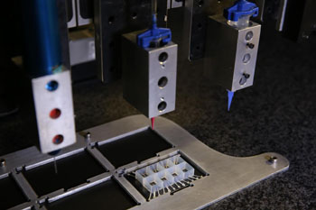
Image: The heart-on-a-chip is made entirely using multi-material three-dimensional printing (3D) in a single automated procedure, integrating six custom printing inks at micrometer resolution (Photo courtesy of Johan Lind, Disease Biophysics Group, and Lori K. Sanders, Lewis Laboratory, Harvard University).
Investigators at Harvard University (Cambridge, MA, USA) recently described a method for fabricating a new class of instrumented cardiac microphysiological devices via multimaterial three-dimensional printing. Specifically, they designed six functional inks, based on piezo-resistive, high-conductance, and biocompatible soft materials that enabled integration of soft strain gauge sensors within micro-architectures that guided the self-assembly of laminar cardiac tissues.
As described in the October 24, 2016, online edition of the journal Nature Materials, the chips contained multiple wells, each with separate tissues and integrated sensors, allowing investigators to study many engineered cardiac tissues at once.
The investigators validated that the embedded sensors provided non-invasive, electronic readouts of tissue contractile stresses inside cell incubator environments. They further applied these devices to study drug responses, as well as the contractile development of human stem cell-derived laminar cardiac tissues over four weeks.
"Our microfabrication approach opens new avenues for in vitro tissue engineering, toxicology, and drug screening research," said senior author Dr. Kit Parker, professor of bioengineering and applied physics at Harvard University. "Translating microphysiological devices into truly valuable platforms for studying human health and disease requires that we address both data acquisition and manufacturing of our devices. This work offers new potential solutions to both of these central challenges."
Related Links:
Harvard University


