Researchers Define the Structure of Parkinson's Disease Protein Aggregates
By LabMedica International staff writers
Posted on 12 Apr 2016
The use of advanced imaging techniques has enabled biochemists to determine the molecular structure of alpha-synuclein protein fibrils such as those found in the brains of individuals with Parkinson's disease.Posted on 12 Apr 2016
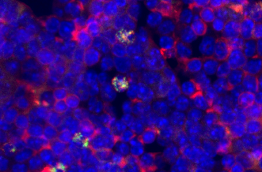
The accumulation of misfolded alpha-synuclein amyloid fibrils leads to the formation of insoluble aggregates that have been implicated in several neurodegenerative diseases, including Parkinson's disease, dementia with Lewy bodies, and Alzheimer's disease. It has been exceedingly difficult to define the structure of alpha-synuclein fibrils due to their insolubility and complexity.
Investigators at the University of Illinois (Champaign-Urbana, USA) and their collaborators used advanced imaging techniques such as magic-angle spinning nuclear magnetic resonance (a type of solid state NMR) to measure the placement of atoms in samples of alpha-synuclein.
They described in the March 28, 2016, online edition of the journal Nature Structural and Molecular Biology a structure with common amyloid features including parallel, in-register beta-sheets and hydrophobic-core residues. The structure revealed substantial complexity arising from diverse structural features including an intermolecular salt bridge, a glutamine ladder, close backbone interactions involving small residues, and several steric zippers stabilizing a new orthogonal Greek-key topology. The results were validated using EM (electron microscope) and X-ray fiber diffraction.
The investigators synthesized alpha-synuclein fibrils according to their structural data and showed that these fibrils induced robust Parkinson's-like pathology in primary neuronal cultures.
"We had to find patterns in the data and systematically test all the possibilities for how the protein would fit together," said senior author Dr. Chad Rienstra, professor of chemistry at the University of Illinois. "It is like when you solve a really complex puzzle, you know you have it right at the end because all the pieces fit together. That is what we got with this structure. This is the first structure of the full-length fibril protein, which is now well established to be important for the pathology of Parkinson's disease. Knowing that structure will open up many new areas of investigation for diagnosing and treating Parkinson's disease."
"We think that the structure that we resolved of alpha-synuclein fibrils will be really significant in the immediate future and has use for diagnosing Parkinson's in patients before they are symptomatic," said Dr. Rienstra. "Once people start having symptoms, whether of Alzheimer's or Parkinson's, in many ways it is a little too late to be effective with therapy. But if you catch it early, I think there is a lot of promise for therapies that are being developed. Those are all relying upon the structures that we are solving."
Related Links:
University of Illinois

Gold Member
Neonatal Heel Incision Device
Tenderfoot


Repetitive Pipette
VWR® Stepper Pro

Immunofluorescence Analyzer
IFA System