Key Finding May Lead to Improved Treatment of Aggressive Brain Cancers
By LabMedica International staff writers
Posted on 20 Jan 2016
Researchers have shown an essential role for the cell microenvironment in the growth of glioblastoma brain cancer cells, a discovery that could lead to a breakthrough in fighting such deadly cancers.Posted on 20 Jan 2016
Stem cells found in the tumors are responsible for making glioblastoma hard to treat because they are drug-resistant and self-renewing. A new study, by researchers at the Institute of Bioengineering and Nanotechnology (IBN; Singapore) of Singapore’s Agency for Science, Technology, and Research (A*STAR), is the first to focus on how the extracellular matrix surrounding the tumor affects development of cancer stem-like cells (CSLCs) in a three-dimensional (3D) microenvironment.

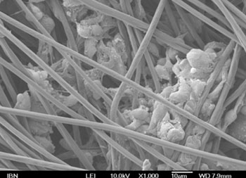
Image: A microscopic image of glioblastoma cells growing along the fibers of the 3D scaffold (Image courtesy of (Photo courtesy of Dr. Andrew Wan laboratory, Institute of Bioengineering and Nanotechnology, Singapore).
“There is currently no cure for glioblastoma, and it is important to eradicate the tumor-initiating cells in order to treat this cancer successfully. By focusing on how the extracellular matrix promotes the development of brain cancer cells, we hope to provide a fresh approach towards tackling the problem, and develop new and more effective therapies,” said IBN executive director Prof. Jackie Y. Ying.
Led by IBN team leader and principal research scientist Dr. Andrew Wan, the researchers studied glioblastoma cell growth in a 3D model using a scaffold of electrospun fibers, compared to in 2D using conventional tissue culture polystyrene plates. The gene and protein expression results showed that the 3D microenvironment promoted the development of brain CSLCs, when compared with the 2D microenvironment.
In particular, they found evidence that two specific types of molecules on the surface of glioblastoma cells, integrin alpha-6 and integrin beta-4, interacted with a specific group of laminin proteins in the extracellular matrix, promoting development of CSLCs. This finding was supported by collaborators at the National Neuroscience Institute using computational approaches to analyze patient tumor and molecular information, which confirmed that these same integrins and laminins were associated with more aggressive brain tumors, particularly grade IV glioblastoma.
“We are excited to have successfully demonstrated that the extracellular matrix and 3D microenvironment work together to affect the stem-like properties in glioblastoma cells. Our finding may also apply to other cell types besides glioblastoma cells, and be used to develop more accurate cancer disease models for drug testing and development. We will conduct further studies with clinical samples from the National Neuroscience Institute, with the goal of improving brain cancer treatment,” said Dr. Wan.
The study, by Ma NKL, Lim JK et al., was published online ahead of print November 26, 2016, in the journal Biomaterials.
Related Links:
Institute of Bioengineering and Nanotechnology


 (3) (1).png)


