Drug Candidate Propels Cancer Cells into Fatal Overdrive
By LabMedica International staff writers
Posted on 23 Aug 2015
A candidate drug that destroys cancer cells by stimulating them to produce more proteins than the cells can actually process was shown to kill a wide variety of cancer cells in culture and to inhibit tumor growth in animal models.Posted on 23 Aug 2015
Investigators at Baylor College of Medicine (Houston, TX, USA) identified the drug MCB-613 as an activator of the steroid receptor coactivators (SRC-1, SRC-2, and SRC-3) while screening a large number of compounds for drugs that would inhibit SRCs. However, when the investigators tested the compound with cultures of cancer cells, they found that MCB-613 could super-stimulate SRCs’ transcriptional activity. Further study revealed that MCB-613 increased SRCs’ interactions with other coactivators and markedly induced ER (endoplasmic reticulum) stress coupled to the generation of toxic reactive oxygen species (ROS).

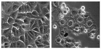
Image: Cancer cells were treated with a control (left) and the overstimulating compound MCB-613 (right) (Photo courtesy of Dr. Lei Wang, Baylor University College of Medicine).
Results published in the August 10, 2015, issue of the journal Cancer Cell revealed that MCB-613 killed human breast, prostate, lung, and liver cancer cells, while sparing normal cells. When administered to 13 mice with breast cancer, MCB-613 reduced tumor growth without causing toxicity, whereas tumors continued to grow by about three-fold over seven weeks in the control group of 14 mice. The toxic effect of the drug was shown to be due to the accumulation of unfolded proteins in the ER. The inability of the ER to cope with such a large number of proteins caused a state of stress to develop that stimulated production of toxic ROS species and the destruction of the cell.
"No prior drug has been previously developed or proposed that actually stimulates an oncogene to promote therapy," said contributing author Dr. David Lonard, associate professor of molecular and cell biology at Baylor College of Medicine. "Our prototype drug works in multiple types of cancers and encourages us that this could be a more general addition to the cancer drug arsenal."
Related Links:
Baylor College of Medicine


