“Nanodaisies” Designed to Transport Drug Cocktail to Cancer Cells
By LabMedica International staff writers
Posted on 10 Jun 2014
The researchers are from the joint biomedical engineering program at North Carolina State University (Raleigh, USA) and the University of North Carolina at Chapel Hill (USA). “We found that this technique was much better than conventional drug-delivery techniques at inhibiting the growth of lung cancer tumors in mice,” stated Dr. Zhen Gu, senior author of the study and an assistant professor in the joint biomedical engineering program. “And based on in vitro tests in nine different cell lines, the technique is also promising for use against leukemia, breast, prostate, liver, ovarian, and brain cancers.”Posted on 10 Jun 2014
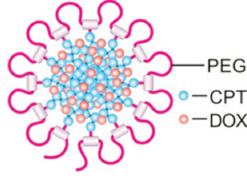
To construct the “nanodaisies,” the researchers started with a solution that contains a polymer called polyethylene glycol (PEG). The PEG forms long strands that have much shorter strands splitting off to either side. Researchers directly attach the anticancer drug camptothecin (CPT) onto the shorter strands and introduce the anticancer drug doxorubicin (Dox) into the solution.

Image: Early tests of the “nanodaisy” drug delivery technique show promise against a number of cancers (Photo courtesy of Ran Mo).
PEG is hydrophilic; CPT and Dox are hydrophobic. As a result, the CPT and Dox cluster together in the solution, wrapping the PEG around themselves. This results in a daisy-shaped drug cocktail, only 50 nm in diameter, which can be injected into a cancer patient. Once injected, the nanodaisies glide through the bloodstream until they are absorbed by cancer cells. In fact, one of the reasons the researchers chose to use PEG is because it has chemical properties that prolong the life of the drugs in the bloodstream.
Once in a cancer cell, the drugs are released. “Both drugs attack the cell’s nucleus, but via different mechanisms,” said Dr. Wanyi Tai, lead author and a former postdoctoral researcher in Dr. Gu’s lab. “Combined, the drugs are more effective than either drug is by itself,” Dr. Gu concluded. “We are very optimistic about this technique and are hoping to begin preclinical testing in the near future.”
The study’s findings were published May 27, 2014, in the journal Biomaterials.
Related Links:
North Carolina State University
University of North Carolina at Chapel Hill


 (3) (1).png)


