Live Imaging Reveals the Brain’s Protein Interactions
By LabMedica International staff writers
Posted on 13 Mar 2014
For the first time, a group of scientists has been able to observe intact interactions between proteins, directly in the brain of a live animal. The new live imaging tool should allow scientists to see the interactions of proteins in the brain of an animal, along diverse points in their natural setting.Posted on 13 Mar 2014
There are more than a trillion cells called neurons that make up a maze of connections in human brains. Each one of these neurons contains millions of proteins that perform different functions. Precisely how individual proteins interact to form the complicated networks of the brain still remains elusive, but is now starting to unravel.
.")
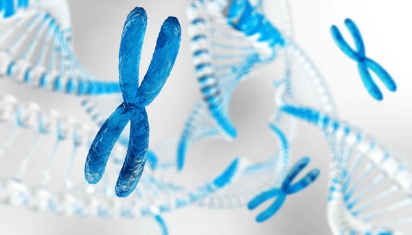
Image: Photonic resonance energy transfer described by Förster, or FRET, occurs when two small proteins come within a very small distance of each other, i.e., 8 nm or less. The fluorescence lifetime of the donor molecule will become shorter—from 3 nanosecond to, possibly, 2.5 nanoseconds. The researchers then interpret this as evidence that the two proteins of interest are physically interacting with each other in a molecular signaling event (Photo courtesy of Akira Chiba/University of Miami).
The new live imaging application was developed by a team of researchers at the University of Miami (UM; FL, USA). “Our ultimate goal is to create the systematic survey of protein interactions in the brain,” said Akira Chiba, professor of biology in the College of Arts and Sciences at UM and lead investigator of the project. “Now that the genome project is complete, the next step is to understand what the proteins coded by our genes do in our body.”
The new technique will allow scientists to visualize the interactions of proteins in the brain of an animal, along different points throughout its development, reported Prof. Chiba, who compares protein interactions to the manner in which organisms associate with each other. “We know that proteins are one billionth of a human in size. Nevertheless, proteins make networks and interact with each other, like social networking humans do,” Prof. Chiba said. “The scale is very different, but it’s the same behavior happening among the basic units of a given network.”
The researchers chose embryos of the fruit fly Drosophila melanogaster as a suitable model for the study. Because of its compact and transparent body, it is possible to see processes inside the Drosophila cells using a fluorescence lifetime imaging microscope (FLIM). The results of the observations are applicable to other animal brains, including the human brain.
The Drosophila embryos in the study contained a pair of fluorescent-labeled proteins: a developmentally essential and ubiquitously present protein called Rho GTPase Cdc42 (cell division control protein 42), labeled with green fluorescent tag and its alleged signaling partner, the regulatory protein WASp (Wiskot-Aldrich syndrome protein), labeled with red fluorescent tag. Combined, these specialized proteins are thought to help neurons grow during brain development. The proteins were chosen because the same (homolog) proteins exist in the human brain as well.
Earlier approaches required chemical or physical treatments that in all probability disrupt or even destroy the cells. That made it impossible to examine the protein interactions in their natural environment. The current study tackles these hurdles by using the occurrence of a phenomenon called Förster resonance energy transfer (FRET). It occurs when two small proteins come within a very small distance of each other (8 nm). The event is interpreted as the time and place where the particular protein interaction occurs within the living animal.
These findings revealed that FRET between the two interacting protein partners happens within neurons, during the time and space that overlaps with the formation of new synapses in the brain of the baby insect. Synapses link up individual neurons in the brain. “Previous studies have demonstrated that Cdc42 and WASp can directly bind to each other in a test-tube, but this is the first direct demonstration that these two proteins are interacting within the brain,” Prof. Chiba concluded.
The researcher’s findings were published February 18, 2014, in the journal PLoS ONE.
Related Links:
University of Miami

Gold Member
Quality Control Material
iPLEX Pro Exome QC Panel


Repetitive Pipette
VWR® Stepper Pro

Hematology Consumables
Bioblood Devices