New Imaging Technology Accelerates Multiple Sclerosis Research
By LabMedica International staff writers
Posted on 09 Jul 2013
Canadian investigators have developed a new magnetic resonance imaging (MRI) technique that detects the characteristic signs of multiple sclerosis in finer detail than ever before, providing a more effective tool for evaluating new treatments. Posted on 09 Jul 2013
The technique analyzes the frequency of electromagnetic waves collected by an MRI scanner, instead of the actual wave size. Although analyzing the number of waves per second had been considered a more sensitive way of identifying changes in tissue structure, the calculations required to generate usable images had been problematic.
.")
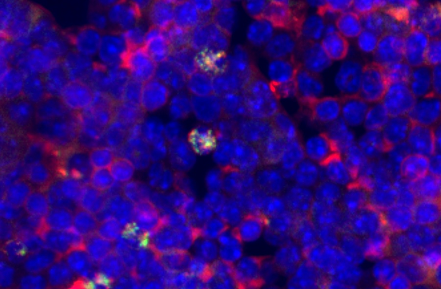
Image: A frequency-based MRI image of an MS patient shows changes in tissue structure (Photo courtesy of the University of British Columbia).
Multiple sclerosis (MS) occurs when an individual’s immune cells attack the protective insulation, known as myelin, which surrounds nerve fibers. The degrading process of myelin hinders the electrical signals transmitted between neurons, resulting in a range of symptoms, including numbness or weakness, vision loss, tremors, fatigue, and dizziness.
Dr. Alexander Rauscher, an assistant professor of radiology, and graduate student Vanessa Wiggerman in the University of British Columbia (UBC) MRI Research Center (Vancouver, BC, Canada), analyzed the frequency of MRI brain scans. With Dr. Anthony Traboulsee, an associate professor of neurology and director of the UBC Hospital MS Clinic, they applied their method to 20 MS patients, who were scanned once a month for six months using both conventional MRI and the new frequency-based method.
Once scars in the myelin (lesions) appeared in conventional MRI scans, Dr. Rauscher and his colleagues went back to earlier frequency-based images of those patients. Looking in the precise areas of those lesions, they found frequency alterations representing tissue damage at least two months before any sign of damage appeared on conventional scans. The results were published according to research published in the June 12, 2013, issue of the journal Neurology, the medical journal of the American Academy of Neurology.
“This technique teases out the subtle differences in the development of MS lesions over time,” Dr. Rauscher concluded. “Because this technique is more sensitive to those changes, researchers could use much smaller studies to determine whether a treatment, such as a new drug, is slowing or even stopping the myelin breakdown.”
Related Links:
University of British Columbia MRI Research Center

Gold Member
Quantitative POC Immunoassay Analyzer
EASY READER+


LAIR2 Antibody Pair Set
LAIR2 Antibody Pair [Biotin]

Chromogenic Culture System
InTray™ COLOREX™ ECC