Single Molecule Visualization Technique Reveals the Structural Dynamics of Telomere Maintenance
By LabMedica International staff writers
Posted on 12 Dec 2012
A recently developed technique for observing single molecules has provided new insights into the structural dynamics involved in the regulation of telomeres.Posted on 12 Dec 2012
A telomere is a region of repetitive nucleotide sequences at each end of a chromosome, which protects the end of the chromosome from deterioration or from fusion with neighboring chromosomes. Telomere regions deter the degradation of genes near the ends of chromosomes by allowing chromosome ends to shorten, which necessarily occurs during chromosome replication. Human telomeres possess a single-stranded DNA (ssDNA) overhang of TTAGGG repeats, which can self-fold into a G-quadruplex structure. Overexpression in cancer cells of the enzyme telomerase, which adds length to telomeres, allows them to divide in perpetuity.
Investigators at the University of Illinois (Urbana; USA) recently developed a single molecule fluorescence assay termed Protein Induced Fluorescence Enhancement (PIFE) and calibrated it on several protein systems. This method circumvented protein labeling and displayed marked distance dependence in the range below four nanometers. The enhancement of fluorescence was based on the photophysical phenomenon whereby the intensity of a fluorophore increased upon proximal binding of a protein. Their data revealed that the method could resolve as little as a single base pair distance at the extreme vicinity of the fluorophore, where the enhancement was maximized.
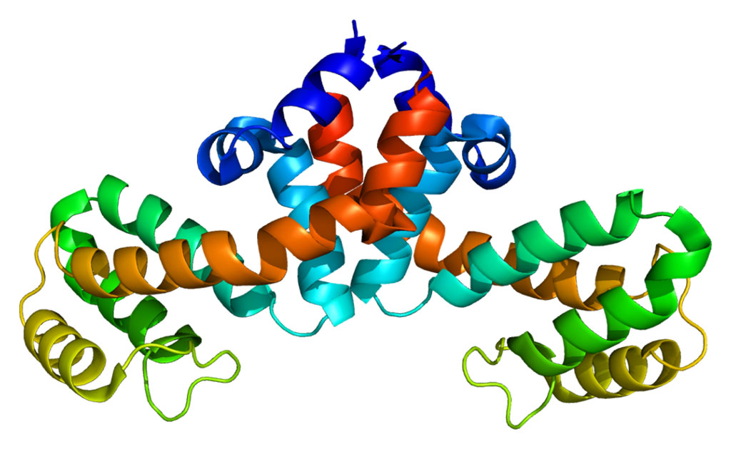
In the current study, the investigators used the PIFE system to analyze the behavior of the enzyme POT1 (protection of telomeres 1), a nuclear protein that functions as a member of a multiprotein complex that binds to the TTAGGG repeats of telomeres, regulating telomere length and protecting chromosome ends from illegitimate recombination, catastrophic chromosome instability, and abnormal chromosome segregation. In particular, they examined the relationship between POT1 and its partner enzyme TPP1 (tripeptidyl-peptidase 1).
They reported in the September 13, 2012, online edition of the journal Structure that POT1 bound specifically to the telomeric overhang and partnered with TPP1 to regulate telomere lengthening and capping. POT1 bound stably to folded telomeric G-quadruplex DNA in a sequential manner, one oligonucleotide/oligosaccharide binding fold at a time. POT1 bound from 3′ to 5′, thereby unfolding the G-quadruplex in a stepwise manner. In contrast, the POT1-TPP1 complex induced a continuous folding and unfolding of the G-quadruplex. The PIFE technique revealed that POT1-TPP1 slid back and forth on telomeric DNA and also on a mutant telomeric DNA to which POT1 could not bind alone.
“Instead of stepwise binding, what we saw was a mobile protein complex, a dynamic sliding motion,” said senior author Dr. Sua Myong, assistant professor of bioengineering at the University of Illinois. “Somehow it was as if the static binding activity of POT-1 is completely lost – the protein complex just slid back and forth. We were able to reproduce the data and confirm it with many different tail lengths of the telomeric DNA, and we know now that the contact between POT-1 and the telomere is somehow altered when the partner protein comes and binds.”
“We are excited about the possibility that this kind of mobility can increase the telomerase extension activity,” said Dr. Myong. “It is somehow engaging the enzyme so that it can stay bound to the DNA longer, so it must involve a direct interaction. We want to extend our basic science knowledge in telomere biology into causes of cancer and we hope that our assay can be useful for telomere-targeted drug screening.”
Related Links:
University of Illinois


 (3) (1).png)


