Differentiated Human Stem Cells Repair Bone Damage in Mouse Model
By LabMedica International staff writers
Posted on 05 Jun 2012
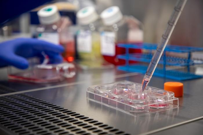
Human-induced pluripotent stem cells (hiPSCs) derived from skin have been cultured in a novel cell and serum-free growth system and then differentiated into mature bone cells that were used to repair damage to bones in a mouse model.Posted on 05 Jun 2012
In 2010, investigators at the University of Michigan (Ann Arbor, USA) described a novel growth system for the culture of hiPSCs. The key to the technique was a synthetic polymer coating, poly[2-(methacryloyloxy)ethyl dimethyl-(3-sulfopropyl)ammonium hydroxide] (PMEDSAH), used in conjunction with a cell and serum-free culture medium.
In the current study, which was published in the May 15, 2012, online edition of the journal Stem Cells, the investigators tested the hypothesis that iPSCs could be maintained in an undifferentiated state in this culture system and subsequently be differentiated into mesenchymal stem cells (iPS-MSCs).
They showed that hiPSCs could be cultured on PMEDSAH and then differentiated into functional MSCs, as confirmed by expression of characteristic MSC biomarkers. To demonstrate the potential of iPS-MSCs to regenerate bone in vivo, the newly derived cells were induced to osteoblast differentiation for four days and then transplanted into immunocompromised mice for eight weeks in order to repair five-millimeter holes in the skulls of the mice. MicroCT (an X-ray computed tomography scanner for small animals) and histologic analyses demonstrated de novo bone formation in the holes in the skulls of animals treated with iPS-MSCs but not for the control group.
By the end of the eight weeks, the mice that had received human-derived bone cells had 4.2 times as much new bone as the controls, as well as the beginnings of marrow cavities. Positive staining for human nuclear antigen and human mitochondria monoclonal antibodies confirmed the participation of the transplanted hiPS-MSCs in the regenerated bone.
"We turn back the clock, in a way," said senior author Dr. Paul Krebsbach, professor of dentistry and biomedical engineering at the University of Michigan. "We are taking a specialized adult cell and genetically reprogramming it, so it behaves like a more primitive cell. The concept is not specific to bone. If we truly develop ways to grow these cells without mouse or animal products, eventually other scientists around the world could generate their tissue of interest."
Related Links:
University of Michigan

Gold Member
Flocked Fiber Swabs
Puritan® Patented HydraFlock®


Hematology Consumables
Bioblood Devices

Food Allergy Screening ELISA Kit
Allerquant 14G B ELISA