Cancer Cells’ DNA Repair Disrupted to Increase Radiation Sensitivity
By LabMedica International staff writers
Posted on 14 Dec 2011
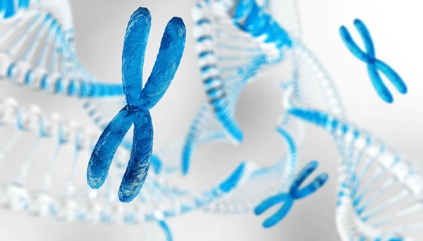
Shortening end caps on chromosomes in human cervical cancer cells disrupts DNA repair signaling, increases the cells’ sensitivity to radiation treatment, and destroys them more rapidly, according to new findings. Posted on 14 Dec 2011
The scientists involved in the study hope to see their laboratory findings, published December 5, 2011, print edition of the journal Cancer Prevention Research, lead to safer, more effective combination therapies for hard-to-treat pediatric brain cancers such as medulloblastoma and high-grade gliomas. To achieve this, they are beginning laboratory tests on brain cancer cells.
“Children with pediatric brain cancers don’t have very many options because progress to find new treatments has been limited the last 30 years,” said Rachid Drissi, PhD, lead investigator on the study and a researcher in the division of oncology from Cincinnati Children’s Hospital Medical Center (OH, USA). “The ability to make cancer cells more sensitive to radiation could allow physicians to use lower radiation doses to lessen side effects. Too many children with brain cancer can develop disabilities or die from treatment.”
Before treating cells with ionizing radiation, the researchers blocked an enzyme called telomerase, found in over 90% of cancer cells but scarcely detectable in most normal human cells. In cancer cells, telomerase helps maintain the length of caps on the ends of chromosomes called telomeres. This helps cancer cells replicate indefinitely, grow, and metastasize, according to Dr. Drissi.
In normal cells lacking the telomerase enzyme, telomeres get shorter each time cells divide. They continue doing so until normal cells stop dividing, reaching a condition called senescence. If this first cell-cycle “stop sign” is bypassed, cells continue dividing until telomeres become critically short and reach a second stopping point, when most cells die. In rare instances, cells sidestep this second “stop sign” and survive. This survival is frequently associated with telomerase activation and the onset of cancer.
This was the foundation for the research Dr. Drissi and his colleagues conducted to compare the radiation sensitivity and survivability of cells based on telomere length. They also monitored DNA repair responses in the cells by looking for specific biochemical signs that indicate whether the repair systems are working.
The tests involved normal human foreskin cells--called fibroblasts--and human cervical carcinoma cells. They exposed the cells to ionizing radiation and studied DNA repair responses as telomeres became increasingly shorter. In the cervical cancer cells, researchers blocked the telomerase enzyme before radiation treatment to induce progressively shorter telomeres.
Both late-stage noncancerous cells with shorter telomeres, and cancer cells with induced shorter telomeres, were more radiosensitive and died more rapidly, according to the study. Among cancer cells with preserved telomere length, close to 10% receiving the maximum dose of ionizing radiation used in the study (8 Gy) survived the treatment. None of the cancer cells with the shortest telomeres survived that exposure.
Researchers reported that the cancer cells became more radiosensitive because material inside the chromosomes--called chromatin--compacted as telomeres became shorter. Compacted chromatin then disrupted the biochemical signaling of a protein called ataxiatelangeietasia mutated (ATM).
ATM is a master regulator of DNA repair and cell division. It sends signals to activate other biochemical targets (H2AX, SMC1, NBS1 and p53) that help direct DNA repair and preserve genetic stability. In telomere-shortened cancer cells, the compacted chromatin inhibited ATM signaling to all of the chromatin-bound targets tested in the study. This disrupted DNA repair responses and increased radiation sensitivity.
The researchers are now assessing their findings in cells from hard-to-treat pediatric brain tumors. These tests begin as Dr. Drissi’s laboratory also leads correlative cancer biology studies of tumor samples from a current clinical trial. The trial is evaluating telomere shortening as a stand-alone therapy for pediatric cancer.
Managed through the US National Institutes of Health’s (Bethesda, MD, USA) Children’s Oncology Group (COG), the multi-institutional phase 1 trial is testing the safety and tumor response capabilities of the drug Imetelstat, which blocks telomerase in cancer cells. Dr. Drissi serves on the clinical trial committee along with Maryam Fouladi, MD, MSc, and medical director of neuro-oncology at Cincinnati Children’s. She leads the medical center's clinical participation in the trial.
Drs. Drissi and Fouladi are starting prep work to develop and seek approvals for a possible clinical trial to evaluate telomere shortening and radiation treatment as a safer, more effective treatment for pediatric brain tumors.
Related Links:
Cincinnati Children’s Hospital Medical Center

Gold Member
Neonatal Heel Incision Device
Tenderfoot


Automatic CLIA Analyzer
Shine i6000

Hematology Consumables
Bioblood Devices