T-cell Activation Independent of Cell Surface Protein Clusters
By LabMedica International staff writers
Posted on 04 Jul 2011
Results from a study conducted with Australia's only super-resolution fluorescence microscope have provided new insights into how immune T-cells become activated to fight infection.Posted on 04 Jul 2011
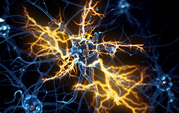
Investigators at the University of New South Wales (Sydney, Australia) used the highly sophisticated super-resolution fluorescence microscope, to observe the movement of Lat (linker for activation of T cells) protein clusters on the surface of T-cells.
The 10-nm resolution obtainable with this instrument revealed that that preexisting Lat domains were neither phosphorylated nor laterally transported to TCR (T cell antigen receptor) activation sites. This finding, which was published in the June 5, 2011, online edition of the journal Nature Immunology, indicated that these clusters did not participate in TCR signaling. Instead, TCR activation resulted in the recruitment and phosphorylation of Lat from subsynaptic vesicles.
Further studies of Lat mutants confirmed that recruitment preceded and was essential for phosphorylation and that both processes were independent of surface clustering of Lat.
"Previously you could see T-cells under a microscope but you could not see what their individual molecules were doing," said senior author Dr. Katharina Gaus, associate professor of vascular research at the University of New South Wales. "Previously it was thought that T-cell signaling was initiated at the cell surface in molecular clusters that formed around the activated receptor. In fact, what happens is that small membrane-enclosed sacks called vesicles inside the cell travel to the receptor, pick up the signal, and then leave again. There is this rolling amplification. The process allows a few receptors to activate a cell and then trigger the entire immune response."
Related Links:
University of New South Wales

New
Gold Member
Neonatal Heel Incision Device
Tenderfoot

New
Total Laboratory Automation Solution
SATLARS Mini T8

New
HIV-1 Molecular Diagnostic Assay
AltoStar HIV RT-PCR Kit 1.5