Protein Structure Revealed by Mass Spectrometry Technique
By LabMedica International staff writers
Posted on 31 May 2011
An advanced mass spectroscopy technique was used to detail the structure of a signaling protein critical to physiological processes involved in major diseases such as diabetes and cancer.Posted on 31 May 2011
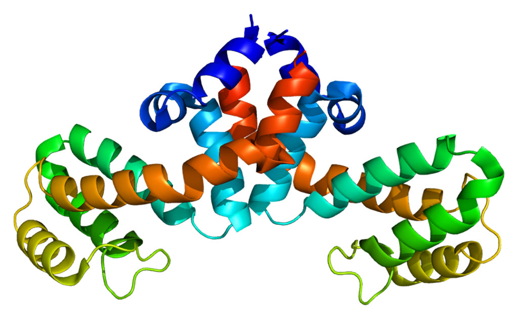
The protein, Epac2 (exchange protein directly activated by cAMP 2), is a guanine nucleotide exchange factor that regulates a wide variety of intracellular processes in response to second messenger cAMP (cyclic adenosine monophosphate).
A collaborative project was carried out by investigators at the University of Texas Medical Branch (Galveston, USA) and the University of California, San Diego (USA) to define the three-dimensional structure of Epac2 in the presence and absence of cAMP using an advanced mass spectroscopy technique known as hydrogen/deuterium exchange mass spectrometry (DXMS).
Results published in the May 20, 2011, issue of the Journal of Biological Chemistry revealed that that cAMP interacted with its two known binding sites on Epac2 in a sequential fashion and that binding of cAMP changed the shape of the protein in a very specific way. This shape change was caused by a major hinge motion centered on the C- terminus of the second cAMP binding domain. This conformational change realigned the regulatory components of Epac2 away from the catalytic core, making the later available for effector binding.
"This study applied a powerful protein structural analysis approach to investigate how a chemical signal called cAMP turns on one of its protein switches, Epac2," said senior author Dr. Xiaodong Cheng, professor of pharmacology and toxicology at the University of Texas Medical Branch.
"DXMS analysis has proved to be an amazingly powerful approach, alone or in combination with other techniques, in figuring out how proteins work as molecular machines, changing their shapes – or morphing – in the normal course of their function," said contributing author Dr. Virgil Woods, professor of medicine at the University of California, San Diego. "This will be of great use in the identification and development of therapeutic drugs that target these protein motions."
Related Links:
University of Texas Medical Branch
University of California, San Diego


