Studies Show Order in Programmed Cell Death
By LabMedica International staff writers
Posted on 02 Apr 2010
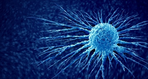
Daily, about 10 billion cells in a human body commit suicide (apoptosis). Cells infected by virus, which are transformed or otherwise dysfunctional, altruistically sacrifice themselves for the greater good. Now, new imaging research has revealed a previously hidden order to this process, showing closely related cells dying in synchrony as a wave of destruction sweeps across their mitochondria, snuffing out the key source of energy that keeps cells alive.Posted on 02 Apr 2010
In experiments published recently in the Journal of Cell Science (November 3, 2010, issue) and the Biophysical Journal (October 21, 2010, issue), researchers in Dr. Sanford M. Simon's laboratory of cellular biophysics at Rockefeller University (New York, NY, USA) photographed the deaths of individual cells, revealing an orderly series of events in the staged shut-down of the cell. The study revealed that the probability of death, as well as the timing, depends on how closely cells are related, not on their proximity to one another or their stage in the cell cycle. The results rule out, for instance, the theory that cells die in a localized cascade accelerated by the secretion of toxic molecules from dying cells nearby.
"What we saw is that, regardless of their location, only the sister cells remained linked in the timing of their deaths,” said Dr. Simon. "It suggests that there is not some nonspecific toxic effect here, but that the variability is in the molecular makeup of the cells--the variability in the population.”
Apoptosis is critical not just in the regular maintenance of life but also in early development--when some cells, such as those that would otherwise form webbing between human fingers, are programmed to die--and in the fine-tuning of the nervous system. "I like to think of it as sculpting, chipping away pieces at a time to create the form,” Dr. Simon noted. A better determination of apoptosis could help clarify certain developmental disorders. Moreover, cell death, or the lack thereof, is important in the pathology of some cancers, in which mutant cells fail to die and grow out of control, forming tumors and metastasizing throughout the body. One potential therapeutic goal would be to learn how to trigger cell death in targeted populations, such as tumors.
Studying the population dynamics of cell death led to the examination, on a much faster timescale, of what was happening inside individual cells during apoptosis. Using single-cell microscopy and fluorescent tags that probe for cell function or for proteins that leave the mitochondria during apoptosis, graduate fellow Patrick Bhola and postdoctoral associate Dr. Alexa Mattheyses captured images as the proteins dispersed through the membrane of one mitochondrion and the process spread in a wave to the other mitochondria in a cell. Some scientists had assumed that this occurred simultaneously to all mitochondria throughout the cell. "This spatial coordination means that there is an upstream signal for release that is spatially localized within individual cells,” stated Dr. Mattheyses.
"The idea in general was to look at individual events in the cells and see if we could get any insights that we could not get looking macroscopically at whole populations of them,” Dr. Simon said. His close-up, observational approach has recently provided new insights into how cells import and export protein cargoes across the cell membrane and how individual HIV particles are created, among other things. Now the microscopy methods are enabling a deeper understanding of apoptosis, stated Dr. Bhola. "It's one of those things where if you can't see what's going on, you tend to assume it's random or all at once,” he said. "But when you get a good look, you find it happens in a very organized fashion.”
Related Links:
Rockefeller University

New
Gold Member
Automatic Hematology Analyzer
CF9600


New
Urine Analyzer
respons® UDS100

New
POC Immunoassay Analyzer
Procise DX