Array Technology Identifies Biologic Components of Liver Disease
By LabMedica International staff writers
Posted on 04 Nov 2009
A combination of bioengineering and medical research has led to a new discovery that could lead the way for more effective treatments for liver disease.Posted on 04 Nov 2009
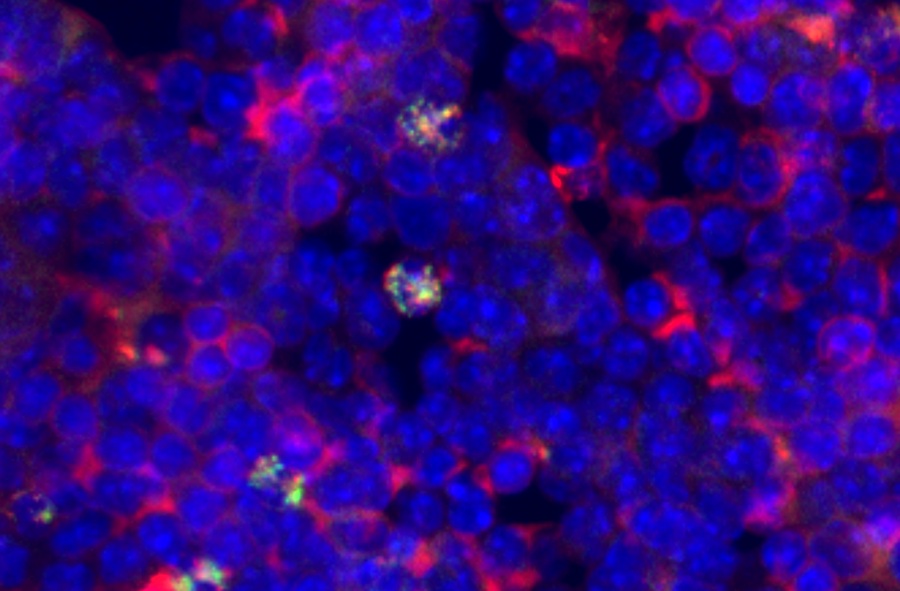
In this study, the researchers, from the University of California, San Diego (UCSD; USA), have utilized an array system that can identify the biologic components that can lead to or alleviate liver disease. The technology works by controlling the range of environments surrounding star-shaped liver cells called hepatic stellate cells (HSCs). HSCs are the major cell type involved in liver fibrosis, which is the formation of scar tissue in response to liver damage. The activated stellate cell is responsible for secreting collagen that produces a fibrous scar, which can lead to cirrhosis.
Current approaches to identify the factors affecting HSC biology typically focus on each factor individually, ignoring the complex crosstalk between the many components acting on the cells. The high-throughput cellular array technology developed by UCSD researchers systematically assesses and probes the complex relationships between hepatic stellate cells and components of their microenvironment. In so doing, they discovered that specific proteins are critical in regulating HSC activation and that the proteins influence one another's actions on the cells. The study's findings were published in the September 2009 issue of the journal Integrative Biology.
"We can spot hundreds of combinations of proteins in the matrix surrounding the cell such as different kinds of collagen--you can spot them individually and in combination, so you can get hundreds of combinations with several proteins,” said Shu Chien, coauthor of the article and bioengineering professor and director of the Institute of Engineering Medicine at UCSD. "We used a spotting pin that's normally used for microarray DNA spotting. But we spot proteins instead of DNA on these slides. "In one step we can look at the physical and chemical microenvironment of the cells,” Dr. Chien added. "Now we can look at the optimum condition that is best for the cells to proliferate and differentiate.”
The high-throughput cellular array technology used in this research was developed by Dr. Chien and his lab colleagues a few years ago chiefly for stem cell research, but it has not been applied to this type of research before involving hepatic stellate cells.
"Our lab is interested in the cells that are responsible for laying down the fibrous scar in all types of chronic liver disease,” said Dr. David Brenner, the dean of the UCSD School of Medicine and coauthor of the study. "In the course of 20 years, people have tried three or four matrixes to try to optimize their growth. Shu Chien mentioned this extracellular matrix array that his lab was using for stem cells, and I thought it would be an incredible opportunity to understand the effects of different matrixes in primary cell cultures. By looking at the array, you get hundreds of different combinations of proteins and you can look at hundreds of cells at once. This will give us new insights for the treatment for liver fibrosis, and it will give us the platform to test new treatments. This also allows us to do the critical experiments that will assess the ability of different drugs to work. There are really no effective therapies for liver fibrosis--there is only transplantation. Any less invasive therapy would be desirable. This is a big advance.”
According to Dr. Chien, such medical developments are the result of the growing synergism between engineering and medicine. This study involves the dissertation work of David Brafman, who received his Ph.D. in bioengineering from UCSD under the joint advisorship of Dr. Chien and Prof. Karl Willert of the department of cellular and molecular medicine. Dr. Chien stated that the next step for this collaborative effort would be to develop a platform that allows scientists and researchers to differentiate cells into the kind they want. It is this type of research, Dr. Chien reported, that gives the field of regenerative medicine a boost. "Eventually we would like to help improve people's health,” he said.
Related Links:
University of California, San Diego

Gold Member
Flocked Fiber Swabs
Puritan® Patented HydraFlock®


Electrolyte Analyzer
BKE-B

Repetitive Pipette
VWR® Stepper Pro