A MicroRNA Pseudo-Oncogene Controls Melanoma Metastasis
By LabMedica International staff writers
Posted on 24 Feb 2009
Cancer researchers have identified a microRNA (miRNA) that functions as a pseudo-oncogene and stimulates the metastasis of melanoma cells.Posted on 24 Feb 2009
Investigators from the New York University (NYU) Medical Center (USA) chose to work on melanoma, since its highly aggressive character makes it an excellent model for probing the mechanisms underlying metastasis, which remains one of the most difficult challenges in treating cancer.
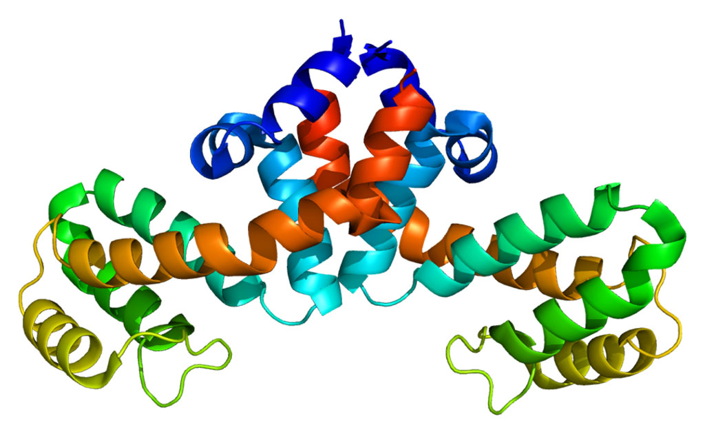
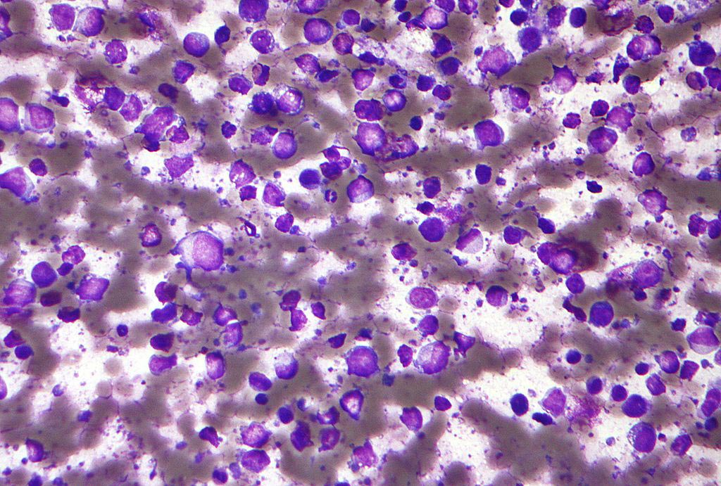
The investigators' work was centered on melanoma cell lines from mouse melanoma models as well as on human cells that were obtained from the large biospecimen bank maintained at the NYU Medical Center. Results published in the February 2, 2009, online edition of the Proceedings of the [U.S.] National Academy of Sciences (PNAS) revealed that the microRNA miRNA182, a member of a miRNA cluster in chromosomal locus (7q31-34) that is frequently amplified in melanoma, was commonly up-regulated in human melanoma cell lines and tissue samples. MiR-182 expression stimulated migration of melanoma cells in vitro and their metastatic potential in vivo, while miR-182 downregulation impeded invasion and triggered apoptosis. Overexpression of miR-182 promoted migration and survival by directly repressing two transcription factors, microphthalmia-associated transcription factor-M (MITF) and FOXO3 (forkhead box O3).
Enhanced expression of either MITF or FOXO3 blocked the pro-invasive effects of miR-182. In human tissues, expression of miR-182 increased with progression from primary to metastatic melanoma and inversely correlated with FOXO3 and MITF levels.
"Melanoma becomes deadly after the cells leave the primary tumor through the blood and metastasize in other organs where they are resistant to therapy,” explained senior author Dr. Eva Hernando, assistant professor of pathology at the New York University Medical Center. "Normal cells are unable to travel and survive in alien locations, so we are very interested in understanding the invasive, adaptive, and resistant traits of the very aggressive melanoma cell.”
"In this study, we began looking at cell lines and then at melanoma tissue,” said Dr. Hernando. "Now that the mechanism has been proven using cell lines and mice, the next step will be to perform in vitro studies with cell lines to assess the effect of anti-miRNA on cell death in both normal and melanoma cells. Once that study is completed, we can use this model for studies in mice to block the growth of the primary melanoma tumor or the metastasis by using anti-miRNA. All these steps will determine if this approach could be eventually applied to humans.”
Related Links:
New York University Medical Center


