Raman Technique Visualizes Details Almost to a Trillionth of a Meter
By Biotechdaily staff writers
Posted on 05 May 2008
A team of researchers has developed a new type of imaging system that can illuminate tumors in living subjects--obtaining images with a precision of nearly one-trillionth of a meter. Posted on 05 May 2008
This technique, called Raman spectroscopy, expands the existing range of tools for the field of molecular imaging, according to team leader Sanjiv Sam Gambhir, M.D., Ph.D., professor of radiology at Stanford University School of Medicine (Stanford, CA, USA). He is the senior author of a study describing the application that was published in the March 31, 2008, advance online issue of the journal Proceedings of the [U.S.] National Academy of Sciences (PNAS).
"This is an entirely new way of imaging living subjects, not based on anything previously used,” said Dr. Gambhir, who directs the Molecular Imaging Program at Stanford. He reported that signals from Raman spectroscopy are stronger and longer lived than other available techniques, and the type of particles used in this method can transmit information about multiple types of molecular targets simultaneously. "Usually we can measure one or two things at a time,” he said. "With this, we can now likely see 10, 20, 30 things at once.
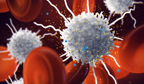
Dr. Gambhir reported that he believes this is the first time Raman spectroscopy has been used to image deep within the body, using tiny nanoparticles injected into the body to serve as beacons. When laser light is beamed from a source outside the body, these specialized particles emit signals that can be measured and translated into a visible indicator of their location in the body.
Postdoctoral scholars Shay Keren, Ph.D., and Cristina Zavaleta, Ph.D., co-first authors of the study, found a way to make Raman spectroscopy a medical application. To accomplish this, they used two types of engineered Raman nanoparticles: gold nanoparticles and single-wall carbon nanotubes. First, they injected mice with the some of the nanoparticles. To see the nanoparticles, they used a special microscope that the group had adapted to view anesthetized mice exposed to laser light. The researchers could see that the nanoparticles migrated to the liver, where they were processed for excretion.
To be able to detect molecular events, according to Dr. Zavaleta, they labeled separate batches of spectrally unique Raman nanoparticles with different "tags” (peptides or antibodies) and then injected them into the body simultaneously to see where they went. For example, if each type of particle migrated to a different tumor site, the newly developed Raman microscope would enable the researchers to separate the signals from each batch of particles.
As part of this proof-of-principle work, Dr. Gambhir's team tagged the gold nanoparticles with different bits of proteins that homed in on different tumor molecules. "We could attach pretty much anything,” said Dr. Gambhir. The Raman effect also lasts indefinitely, so the particles do not lose effectiveness as indicators as long as they stay in the body.
Using a microscope they modified to detect Raman nanoparticles, the researchers were able to visualize targets on a scale 1,000 times smaller than what is now obtainable by the most precise fluorescence imaging using quantum dots. When modified for human use, they researchers reported, the technique has the potential to be useful during surgery, for example, in the removal of cancerous tissue. The extreme sensitivity of the imager could enable detection of even the smalltest malignant tissues.
A clinical trial is planned to test the gold nanoparticles in humans for possible use in conjunction with a colonoscopy to indicate early-stage colorectal cancer.
Related Links:
Stanford University School of Medicine

Gold Member
Quantitative POC Immunoassay Analyzer
EASY READER+


New
Multi-Chamber Washer-Disinfector
WD 390

New
HPV Test
Allplex HPV28 Detection