X-Ray Crystallography Reveals How Substances Enter Cells
By Biotechdaily staff writers
Posted on 11 Dec 2007
A team of researchers has captured a vital step in the metabolic mechanisms that allow substances, such as nutrients and drug treatments, to travel in and out of cells. Posted on 11 Dec 2007
A research team led by Dr. Jue Chen, an associate professor of biological sciences at Purdue University (West Lafayette, IN, USA), obtained a snapshot of the small protein gate complex that opens and closes pathways through the protective cellular membrane. The gates, regulated by small protein units that push them open and closed, bring nutrients into the cell and flush out waste.
The Purdue-led team was the first to achieve an image of the middle step of the process, capturing the molecular interactions as material passes through the membrane. "By understanding the mechanisms of this process, researchers may be able to design more effective treatments for diseases that involve this group of proteins, such as cancer and cystic fibrosis,” said Dr. Chen, who also is a member of Purdue's structural biology group within the College of Science. "With this knowledge, researchers may be able to inhibit or activate this mechanism, depending on what is needed to counteract the disease. For instance, many cancer cells are resistant to drug treatments because the cells pump the drugs out through these channels before they can work.”
Dr. Amy Davidson, an associate professor of chemistry at Purdue and collaborator on this work with Dr. Chen, reported that capturing an image of the intermediate stage is a huge step toward learning the complete process. "If you look at only the before and after stages, you don't really know all that goes on,” said Dr. Davidson. "The intermediate stage provides all of this information about how the process really works. It shows all of the main components of the system interacting, which had not been seen before. It is a snapshot of what happens halfway through the entry process and is a very clear picture of how things work.”
The investigators utilized X-ray crystallography to obtain an image of a particular protein, called an ABC (adenosine triphosphate- [ATP]-binding cassette) transporter protein, as it moved material through the cellular membrane. The study was published in the November 22, 2007, issue of the journal Nature.
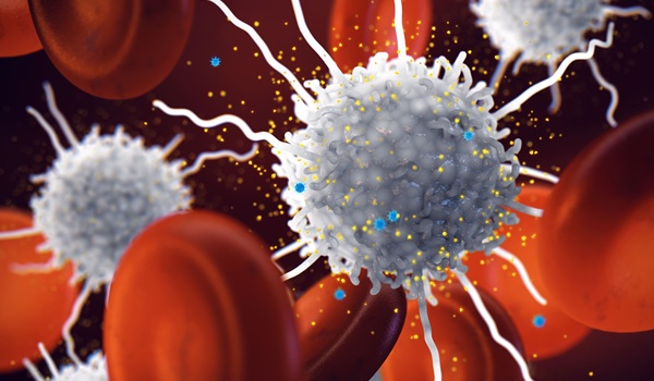
ABC proteins are present in every living thing and have important biologic functions. Scientists have identified 49 different ABC proteins in humans and have found that more than a dozen disease states are associated with malfunctions of these proteins, including macular dystrophy and abnormalities in the regulation of cholesterol and insulin secretion.
Dr. Chen's team isolated ABC proteins from an Escherichia coli bacterium, which is the standard research subject for this field of work. The ABC proteins are structurally very similar to those in human cells, and most of the principles can be directly applied to humans, according to Dr. Chen.
Membrane proteins are extremely difficult to study, according to Dr. Chen. Whereas most proteins dissolve in water and can be easily crystallized and examined, membrane proteins dissolve only in fatty substances, making it hard to isolate them for study. The collaboration between Drs. Chen and Davidson, a structural biologist and a biochemist, was the key to success in capturing the intermediate structure, according to Dr. Chen. Dr. Davidson identified a particular mutant of the ABC protein that locks halfway through the process. The mutant trapped the protein complex in a stable form that allowed Dr. Chen to crystallize and visualize the structure.
"We have the common goal to learn how this family of proteins works, but we take different approaches,” Dr. Chen said. "It is important to communicate with scientists in other fields who can offer clues and tools to help reach your goal. We used genetic data and biochemical data to learn what we needed to crystallize and solve the structure.”
Whereas structural biologists attempt to obtain images of a protein as it performs a process, biochemists utilize indirect methods to understand the nature of a protein and how it moves through different conformations. "Collaboration allows us to marry visual structures to other techniques that let you see the motions,” Dr. Davidson noted. "It all came together beautifully and matched the model we proposed in 2001 of what things should look like during this process. Through genetic and biochemical work, we determined which proteins were important to this process, but we didn't know exactly how they worked. The image of the structure answers these questions and clearly shows the specific interactions.”
In the next phase of the study, the researchers will try to determine the structure of another conformation of the protein that explains the other half of the process: how material is delivered into the cell.
Related Links:
Purdue University

New
Gold Member
Automatic Hematology Analyzer
CF9600


New
CMV CLIA Diagnostic
CLIA CMV IgA Screen Group

New
Automated Urinalysis Solution
UN-9000