New Target for Anti-Cancer Therapies Provided by Telomerase Enzyme Structure
By Biotechdaily staff writers
Posted on 03 Dec 2007
Posted on 03 Dec 2007
.")
Computer artwork of two chromosomes highlighted where the telomeres are located. Telomeres consist of short, repeated sequences of DNA and proteins that play an important role in cellular aging (Photo courtesy of Hybrid Medical Animation / SPL).
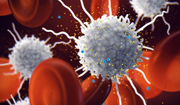
Inappropriate activation of a single enzyme, telomerase, is associated with the uncontrollable proliferation of cells seen in as many as 90% of all of human cancers. Since the mid-1990s, when telomerase was first detected in human tumors, scientists have looked at the enzyme as an ideal target for developing widely effective anti-cancer drugs.
The researchers, from the Wistar Institute (Philadelphia, PA, USA), published their findings November 13, 2007, in the journal Structure, and reported that their research may help scientists develop strategies to devise the first direct inhibitors of telomerase. Telomerase also has been shown to play a critical role in normal aging, and the new study may provide insights into that vital life process as well. The potential for creating new cancer treatments, however, is the most important immediate implication of the study.
"Knowing the physical structure of this complex will give pharmaceutical companies a direct target for designing drugs that disrupt a mechanism that telomerase uses to assemble itself,” stated Emmanuel Skordalakes, Ph.D., an assistant professor in the Gene Expression and Regulation Program at Wistar, and senior author on the study. "Such drugs could well have significant anti-cancer activity.”
Telomerase is necessary for normal cell division and survival, and has been associated with aging and cancer. In humans, the typical role of telomerase is to add multiple repeats of a short length of DNA to the ends of chromosomes, known as telomeres, thereby preventing damage and the loss of genetic information during DNA replication. It performs this vital role in developing embryos and in a few specialized cell lines, including stem cells.
In normal adult cells, however, telomerase is turned off almost entirely to prevent the hazards of runaway cell proliferation. This lack of telomerase activity is also associated with normal aging and underlies a seminal observation known as the Hayflick limit. At Wistar in the 1960s, Leonard Hayflick, Ph.D., noted that cells in culture divide only approximately 50 times before dying. Later on, scientists tied this effect to the shortening of telomeres with each cell division when telomerase is no longer active in the cell.
Cancer cells, however, frequently regain the ability to produce telomerase, permitting them to replicate indefinitely. Although scientists have looked for ways to inhibit this enzyme, a lack of detailed information on the enzyme's structure has hindered progress.
Earlier research has shown that telomerase is comprised of multiple protein components and a length of RNA that is used as a template to create the short DNA repeats that are added to the ends of chromosomes. For telomerase to function, the RNA and protein components of telomerase must interact to form a stable complex capable of DNA replication. This interaction occurs mainly on the so-called TRBD domain, which plays a fundamental role in complex formation and full assembly of the enzyme.
"Studies show if you delete the TRBD domain from telomerase, the enzyme is inactive because it can no longer assemble with RNA,” Dr. Skordalakes noted. "Without the RNA, the enzyme can no longer replicate telomeres.”
To obtain a clear view of this interaction, Dr. Skordalakes and co-workers obtained the three-dimensional structure of TRBD using X-ray crystallography, a technique that analyzes the diffraction patterns of X-rays beamed at crystals of a molecule to determine the molecule's atomic structure. The investigators' research revealed that the TRBD domain is shaped like a boomerang, with a structural organization that leads to the formation of a narrow well-defined pocket on the surface of the protein that enables the enzyme to bind the single-stranded RNA used as a template for the DNA repeats.
A second RNA-binding site is formed by a large cavity that serves as an extension of the single-stranded RNA-binding pocket. The degree of these RNA interactions indicates the important role of this domain in stabilizing the complex, according to Dr. Skordalakes.
Related Links:
Wistar Institute

New
Gold Member
Aspiration System
VACUSAFE


New
Automatic CLIA Analyzer
Shine i6000

New
HPV Molecular Test
BD Onclarity HPV Assay