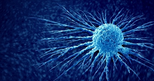
Cancer Identified By Scanning Surface Veins with Lasers
By Biotechdaily staff writers
Posted on 17 Sep 2007
A new technology for cancer detection that eliminates the need for drawing blood has recently been developed.Posted on 17 Sep 2007
Researchers from the University of Purdue (West Lafayette, IN, USA) collaborated with cancer and biotechnology specialists from the Mayo Clinic (Rochester, MN, USA) to develop technology to detect tumor cells within the human body. By shining a laser on surface veins, such as those on the wrist and inside the cheek, researchers are able to reveal and count circulating tumor cells.
Optical imaging provides high resolution and chemical specificity for cancer detection, but it typically is limited from limited penetration depth, making it hard to reach tumors inside the body, according to Dr. Ji-Xin Cheng, an assistant professor of chemistry and biomedical engineering at Purdue.
The technique could provide clinicians and patients results in minutes, and save the medical industry millions of dollars in testing equipment, according to Wei He, a graduate student in the Purdue department of chemistry and the department of biomedical engineering. He worked on the project with Drs. Low and Cheng. By directly labeling tumor cells while they are in the bloodstream, some of the costs and problems associated with testing drawn blood samples can be avoided, according to Mr. He.
An article describing the technology and detection technique was published in the July 10, 2007, issue of the journal Proceedings of the [U.S.] National Academy of Sciences. The technique uses a fluorescent tumor-specific probe that labels tumor cells in circulation. When hit by a laser, which scans across the diameter of the blood vessel 1,000 times per second, the tumor cells glow and become visible. The in-vivo flow detection was performed on a two-photon fluorescence microscope in Dr. Cheng's lab. The researchers compared several methods and found two-photon fluorescence provides the best signal to background ratio. The technology is able to scan every cell that is pumped through the vessel.
The researchers have developed two labeling agents that attach to different forms of cancer. One label targets ovarian, non-small lung, kidney, and endometrial cancer, and the other targets prostate cancer. These labels would be administered through an injection. The first label has already been evaluated in humans and has no adverse side effects and could potentially be administered weekly.
The laser penetrates to a depth of 100 microns and is able to examine shallow blood vessels near the surface of the skin. Sophisticated optical technology could be incorporated into the technology platform and enable the method to reach deeper vessels that handle larger volumes of blood, according to Dr. Cheng.
Related Links:
University of Purdue
Mayo Clinic

Gold Member
Flocked Fiber Swabs
Puritan® Patented HydraFlock®


New
Pipette Calibration System
Artel PCS®

New
Steam Sterilizer
Hi Vac II Line