AI Bone Marrow Mapping Provides New Tool to Track Blood Cancer Severity
Posted on 21 Jul 2026
Myelodysplastic neoplasms, a group of blood cancers that primarily affect older adults, are challenging to stage and monitor, often requiring repeated bone marrow biopsies that can yield uncertain interpretations. The disease can progress to acute myeloid leukemia, highlighting the need for objective, reproducible risk measures. Conventional histopathology and genetic testing can also leave a diagnostic gray zone. New findings demonstrate an artificial intelligence approach that turns routine marrow images into a quantitative disease score.
Weill Cornell Medicine investigators developed the MDS-Microarchitectural Perturbation Score (MDS-MAPS), an artificial intelligence (AI) method that quantifies disease severity in bone marrow biopsies from patients with myelodysplastic neoplasms (MDS). The approach compares the spatial positions and morphometric characteristics of every hematopoietic cell in a patient sample with those in healthy marrow to generate a single score. It analyzes digital images produced through routine staining and imaging workflows commonly available in hospital pathology laboratories.
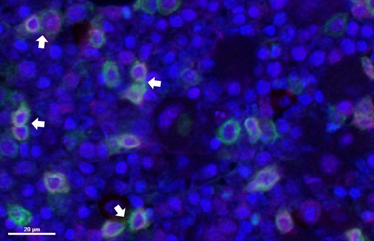
MDS-MAPS ranks samples using 82 features associated with normal tissue and genetic MDS subtypes. The investigators used fully deidentified patient samples and indicate that the score can be generated at diagnosis and tracked longitudinally. Lower scores resemble healthy tissue, while higher scores reflect greater disease-related architectural disruption.
In computer modeling, the tool classified disease status more accurately than current assessment methods. Beyond scoring, the study found that healthy bone marrow follows a conserved architecture and that stem cells become increasingly misplaced as MDS advances. Reduced expression of the CXCR4 receptor was observed in stem cells from patients, particularly when TP53 mutations were present, offering insights that could further refine pathology monitoring.
The work was published June 30 in Leukemia. The team plans to validate the approach in larger cohorts and evaluate it in additional MDS forms and increasingly recognized precursor conditions.
“Using AI to assist us in looking at the spatial architecture of the bone marrow using widely available laboratory assays allows us to improve our capability to assess patients’ prognosis and perhaps triage them for precision therapies,” said Dr. David Redmond, assistant professor of computational biology research in medicine at Weill Cornell Medicine.
“We can generate an MDS-MAPS value for a patient at diagnosis and track how it changes over time,” said Dr. Sanjay Patel, clinical chief of hematopathology and associate professor of pathology and laboratory medicine at Weill Cornell Medicine. “It distills something very complex down into a numerical value. It is easier for a patient to understand whether their score is trending in the right or wrong direction, or whether their disease is more likely stable.”
Related Links
Weill Cornell Medicine