Imaging Platform Maps Lipid Accumulations in Fabry Heart Tissue
Posted on 16 Jul 2026
Mapping the spatial distribution of disease-relevant molecules within tissue remains a diagnostic challenge, particularly before alterations are visible by conventional microscopy. In Fabry disease, a rare genetic metabolic disorder, globotriaosylceramides (Gb3) accumulate in organs including the heart and kidneys, driving progressive damage. Precise localization of these lipids could inform more reliable assessments of affected tissue. Researchers now demonstrate a correlative imaging approach that delineates lipid distributions in a cardiac model of Fabry disease.
Investigators at the Leibniz Institute for Analytical Sciences (ISAS; Dortmund, Germany) combined Raman microscopy with AP-MALDI-MSI (Atmospheric-Pressure Matrix-Assisted Laser Desorption/Ionization Mass Spectrometry Imaging) to generate a high-resolution molecular map of tissue. The work is described as the first integration of these complementary modalities on the same tissue section for this purpose. By combining both techniques, the approach visualizes multiple biomolecular classes simultaneously and reveals their spatial organization.
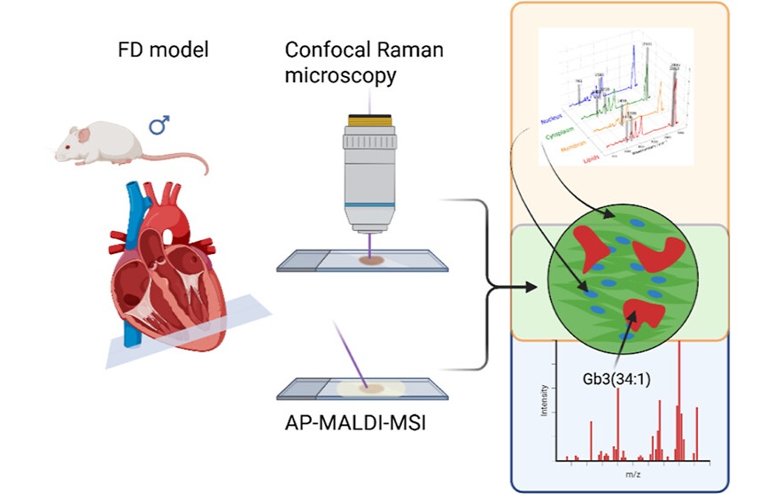
Raman microscopy captures chemical signatures of molecular classes and tissue structure with pixel sizes down to approximately two micrometers, while AP-MALDI-MSI identifies and localizes individual molecules with pixels as small as five micrometers. Specially developed software automatically merges the two datasets and aligns them on the same tissue section with micrometer-level precision.
As an application example, the team analyzed heart tissue from Fabry disease mouse models. Automatic co-registration aligned the Raman and AP-MALDI-MSI images with pixel-perfect precision, enabling spatially resolved assessment of disease-relevant lipids within the myocardium.
The resulting molecular map showed that Gb3 is not uniformly distributed across cardiac tissue. Instead, distinct molecular variants of Gb3 formed minute, well-defined accumulations that could be resolved at micrometer scale within affected regions.
The development and case study were published in Analytical Chemistry on June 4, 2026. The researchers next plan to apply the method to patient tissue to better elucidate disease mechanisms in individual manifestations of Fabry disease, with the goal of enabling new approaches to diagnosis and treatment. They also note potential relevance for other conditions characterized by local molecular changes in tissue, including cardiovascular and metabolic diseases.
“Only by combining Raman microscopy with mass spectrometry imaging is it possible to obtain a comprehensive picture of the molecular processes within the tissue. For a reliable diagnostic assessment, it's important to know exactly where in the tissue Gb3 molecules accumulate,” said Prof. Dr. Sven Heiles, head of the lipidomics junior research group at ISAS and one of the corresponding authors.
“The genetic cause of Fabry disease and elevated Gb3 levels in the blood have long been known, and typical target organs with lipid storage have been described in studies. However, the distribution of lipids in human tissue at the cellular and subcellular levels—as well as the associated interindividual heterogeneity—had previously been inadequately characterized. These new findings could help us better understand the course of the disease in the future,” said Prof. Dr. Kristina Lorenz, head of the Translational Research Department at ISAS and one of the corresponding authors.
Related Links
Leibniz Institute for Analytical Sciences