Manual Aspiration by Monovette Reduces Hemolytic Sampling
|
By LabMedica International staff writers Posted on 18 Aug 2021 |

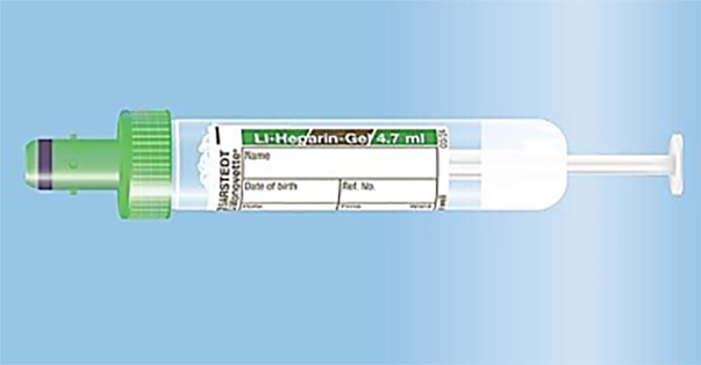
Image: The manual aspiration of the 4.7 mL S-Monovette Lithium-Heparin Gel tube reduces hemolytic sampling (Photo courtesy of SARSTEDT AG)
Hemolytic blood samples are the number one cause for specimen rejection at emergency departments (ED). Triggered by unsuitable blood sampling material or incorrect handling and a related strong vacuum force, hemolytic samples often must be retaken.
Hemolysis refers to the release of hemoglobin and other intracellular components into the surrounding plasma due to the ruptured cell membrane of erythrocytes. The root cause of hemolysis in vitro is an improper sample drawing or, more specifically, an evolving strong vacuum force.
Clinical Laboratorians at the HFR Fribourg-Hôpital Cantonal (Villars-sur-Glâne, Switzerland) conducted a head-to-head study between January and April 2019. In the first eight weeks, all specimens were collected using BD Vacutainer Lithium-Heparin Gel tubes (Vacutainer, Becton, Dickinson and Company, Franklin Lakes, NJ, USA), in the second eight weeks, blood was taken using S-Monovette Lithium-Heparin Gel tubes (SARSTEDT AG, Nümbrecht, Germany) in aspiration mode. Specimens were categorized into five classes (0–30, 31–50, 51–75, 76–100, and 101+ mg/dL of cell-free hemoglobin) and for the statistical analyses, all samples exceeding 30 mg/dL were classified as hemolytic.
All blood samples from the emergency department were evaluated using a Cobas 6000, (Roche Diagnostics, Rotkreuz, Switzerland) a state-of-the-art analyzing system, and their Hemolysis Index (HI 1 = 1 mg of free cell hemoglobin in 1 dL blood plasma) was determined. Data were collected on 4,794 blood specimens (Vacutainer: 2,634 samples, S-Monovette: 2,160 samples).
The scientists reported that overall, 11.3 % of samples were rated as hemolytic because their concentration of hemolysis exceeded 30 mg/dL. This proportion differed considerably between specimens drawn by Vacutainer (17.0 %) and S-Monovette (4.3 %), meaning that, in proportion, there were four times as many hemolytic samples when using Vacutainer. While the percentage of non-hemolytic samples (HI of 0–30 mg/dl) was substantially higher for specimens drawn by S-Monovette (95.7 %) than Vacutainer (83.0 %), the opposite was true for all HI categories above 30 mg/dl.
The authors concluded that regarding hemolysis rates, a slow manual aspiration using S-Monovette was superior to vacuum tubes with predefined filling volumes, as demonstrated in the setting of their ED, which has important practical implications. This blood sampling process could be highly beneficial, not only from a financial point of view, but also with regards to reducing unnecessary tasks and stress for nursing staff and improving patient outcome overall. The study was published on July 28, 2021 in the journal Practical Laboratory Medicine.
Related Links:
HFR Fribourg-Hôpital Cantonal
Becton, Dickinson and Company
SARSTEDT AG
Roche Diagnostics
Latest Technology News
- New Diagnostic System Achieves PCR Testing Accuracy
- DNA Biosensor Enables Early Diagnosis of Cervical Cancer
- Self-Heating Microfluidic Devices Can Detect Diseases in Tiny Blood or Fluid Samples
- Breakthrough in Diagnostic Technology Could Make On-The-Spot Testing Widely Accessible
- First of Its Kind Technology Detects Glucose in Human Saliva
- Electrochemical Device Identifies People at Higher Risk for Osteoporosis Using Single Blood Drop
- Novel Noninvasive Test Detects Malaria Infection without Blood Sample
- Portable Optofluidic Sensing Devices Could Simultaneously Perform Variety of Medical Tests
- Point-of-Care Software Solution Helps Manage Disparate POCT Scenarios across Patient Testing Locations
- Electronic Biosensor Detects Biomarkers in Whole Blood Samples without Addition of Reagents
- Breakthrough Test Detects Biological Markers Related to Wider Variety of Cancers
- Rapid POC Sensing Kit to Determine Gut Health from Blood Serum and Stool Samples
- Device Converts Smartphone into Fluorescence Microscope for Just USD 50
- Wi-Fi Enabled Handheld Tube Reader Designed for Easy Portability
Channels
Clinical Chemistry
view channel
3D Printed Point-Of-Care Mass Spectrometer Outperforms State-Of-The-Art Models
Mass spectrometry is a precise technique for identifying the chemical components of a sample and has significant potential for monitoring chronic illness health states, such as measuring hormone levels... Read more.jpg)
POC Biomedical Test Spins Water Droplet Using Sound Waves for Cancer Detection
Exosomes, tiny cellular bioparticles carrying a specific set of proteins, lipids, and genetic materials, play a crucial role in cell communication and hold promise for non-invasive diagnostics.... Read more
Highly Reliable Cell-Based Assay Enables Accurate Diagnosis of Endocrine Diseases
The conventional methods for measuring free cortisol, the body's stress hormone, from blood or saliva are quite demanding and require sample processing. The most common method, therefore, involves collecting... Read moreMolecular Diagnostics
view channel
Blood Test Accurately Predicts Lung Cancer Risk and Reduces Need for Scans
Lung cancer is extremely hard to detect early due to the limitations of current screening technologies, which are costly, sometimes inaccurate, and less commonly endorsed by healthcare professionals compared... Read more
Unique Autoantibody Signature to Help Diagnose Multiple Sclerosis Years before Symptom Onset
Autoimmune diseases such as multiple sclerosis (MS) are thought to occur partly due to unusual immune responses to common infections. Early MS symptoms, including dizziness, spasms, and fatigue, often... Read more
Blood Test Could Detect HPV-Associated Cancers 10 Years before Clinical Diagnosis
Human papilloma virus (HPV) is known to cause various cancers, including those of the genitals, anus, mouth, throat, and cervix. HPV-associated oropharyngeal cancer (HPV+OPSCC) is the most common HPV-associated... Read moreHematology
view channel
Next Generation Instrument Screens for Hemoglobin Disorders in Newborns
Hemoglobinopathies, the most widespread inherited conditions globally, affect about 7% of the population as carriers, with 2.7% of newborns being born with these conditions. The spectrum of clinical manifestations... Read more
First 4-in-1 Nucleic Acid Test for Arbovirus Screening to Reduce Risk of Transfusion-Transmitted Infections
Arboviruses represent an emerging global health threat, exacerbated by climate change and increased international travel that is facilitating their spread across new regions. Chikungunya, dengue, West... Read more
POC Finger-Prick Blood Test Determines Risk of Neutropenic Sepsis in Patients Undergoing Chemotherapy
Neutropenia, a decrease in neutrophils (a type of white blood cell crucial for fighting infections), is a frequent side effect of certain cancer treatments. This condition elevates the risk of infections,... Read more
First Affordable and Rapid Test for Beta Thalassemia Demonstrates 99% Diagnostic Accuracy
Hemoglobin disorders rank as some of the most prevalent monogenic diseases globally. Among various hemoglobin disorders, beta thalassemia, a hereditary blood disorder, affects about 1.5% of the world's... Read moreImmunology
view channel
Diagnostic Blood Test for Cellular Rejection after Organ Transplant Could Replace Surgical Biopsies
Transplanted organs constantly face the risk of being rejected by the recipient's immune system which differentiates self from non-self using T cells and B cells. T cells are commonly associated with acute... Read more
AI Tool Precisely Matches Cancer Drugs to Patients Using Information from Each Tumor Cell
Current strategies for matching cancer patients with specific treatments often depend on bulk sequencing of tumor DNA and RNA, which provides an average profile from all cells within a tumor sample.... Read more
Genetic Testing Combined With Personalized Drug Screening On Tumor Samples to Revolutionize Cancer Treatment
Cancer treatment typically adheres to a standard of care—established, statistically validated regimens that are effective for the majority of patients. However, the disease’s inherent variability means... Read moreMicrobiology
view channel
New CE-Marked Hepatitis Assays to Help Diagnose Infections Earlier
According to the World Health Organization (WHO), an estimated 354 million individuals globally are afflicted with chronic hepatitis B or C. These viruses are the leading causes of liver cirrhosis, liver... Read more
1 Hour, Direct-From-Blood Multiplex PCR Test Identifies 95% of Sepsis-Causing Pathogens
Sepsis contributes to one in every three hospital deaths in the US, and globally, septic shock carries a mortality rate of 30-40%. Diagnosing sepsis early is challenging due to its non-specific symptoms... Read morePathology
view channelAI-Powered Digital Imaging System to Revolutionize Cancer Diagnosis
The process of biopsy is important for confirming the presence of cancer. In the conventional histopathology technique, tissue is excised, sliced, stained, mounted on slides, and examined under a microscope... Read more
New Mycobacterium Tuberculosis Panel to Support Real-Time Surveillance and Combat Antimicrobial Resistance
Tuberculosis (TB), the leading cause of death from an infectious disease globally, is a contagious bacterial infection that primarily spreads through the coughing of patients with active pulmonary TB.... Read moreIndustry
view channel
ECCMID Congress Name Changes to ESCMID Global
Over the last few years, the European Society of Clinical Microbiology and Infectious Diseases (ESCMID, Basel, Switzerland) has evolved remarkably. The society is now stronger and broader than ever before... Read more
Bosch and Randox Partner to Make Strategic Investment in Vivalytic Analysis Platform
Given the presence of so many diseases, determining whether a patient is presenting the symptoms of a simple cold, the flu, or something as severe as life-threatening meningitis is usually only possible... Read more
Siemens to Close Fast Track Diagnostics Business
Siemens Healthineers (Erlangen, Germany) has announced its intention to close its Fast Track Diagnostics unit, a small collection of polymerase chain reaction (PCR) testing products that is part of the... Read more


.jpg)

