PCR Method Identifies Microsatellite Instability in Tumor-Derived Samples
|
By LabMedica International staff writers Posted on 31 Mar 2021 |

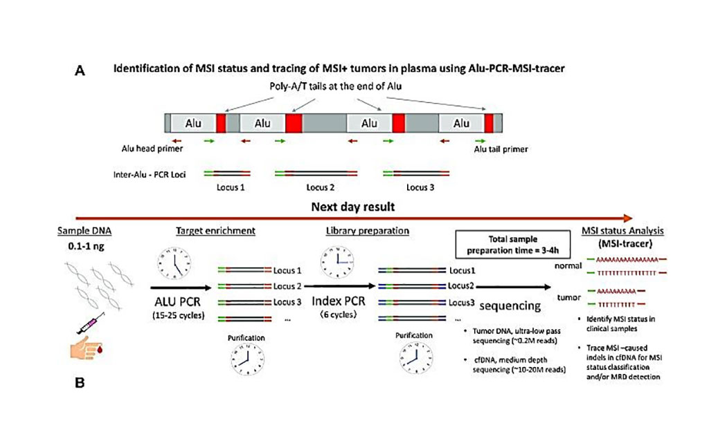
Image: Schematic diagram for the identification of MSI status and tracing MSI+ tumors in plasma using Alu-PCR-MSI tracer (Photo courtesy of Dana-Farber Cancer Institute)
Sensitive detection of microsatellite instability (MSI) in tissue or liquid biopsies using next generation sequencing (NGS) has growing prognostic and predictive applications in cancer. However, the complexities of NGS make it cumbersome as compared to established multiplex-PCR detection of MSI.
Tumors with MSI accumulate high numbers of somatic microsatellite (MS) insertions or deletions (indels), due to a loss of normal mismatch repair (MMR) ability. High levels of MSI are predictive for colorectal cancer (CRC) therapy outcome in chemotherapy and immunotherapy and has been associated with distinct characteristics and favorable results including better prognosis, a higher 5-year survival, and lesser metastasis.
Radiation Oncologists at the Dana-Farber Cancer Institute (Boston, MA, USA) and their colleagues obtained snap-frozen colon adenocarcinoma stage II/III and paired normal tissue biopsies from treatment-naïve patients were obtained from the Massachusetts General Hospital Tumor Bank and gDNA was extracted using the Blood and Tissue kit (Qiagen, Hilden, Germany). Plasma was acquired from healthy volunteers and from stage I/II colon adenocarcinoma treatment-naïve patients. cfDNA was isolated using Qiagen’s QIAamp Circulating Nucleic Acids Kit. The concentration of isolated DNA was quantified on a Qubit 3.0 fluorometer using dsDNA HS assay kit (Thermo Fisher Scientific, Waltham, MA, USA).
To detect MSI the team developed a method called inter-Alu-PCR followed by targeted NGS that combines the practical advantages of multiplexed-PCR with the breadth of information provided by NGS. Inter-Alu-PCR employs poly-adenine repeats of variable length present in every Alu element and provides a massively-parallel, rapid approach to capture poly-A-rich genomic fractions within short 80–150bp amplicons generated from adjacent Alu-sequences. A custom-made software analysis tool, MSI-tracer, enables Alu-associated MSI detection from tissue biopsies or MSI-tracing at low-levels in circulating-DNA.
To establish the method's limit of detection in tissue samples, the scientists tested multiple scenarios using serial dilutions of MSI-H tumor DNA from colon cancer into matched normal DNA. The team used droplet digital PCR to validate the dilution approach using tumor-specific somatic mutations such as KRAS for a subset of the mutations. When a paired normal sample was not present, the team found that that the method had a limit of detection of 0.15% to 0.5% percent for somatic indels using a low-tumor purity clinical sample. When matched normal tissue was available, inter-Alu-PCR had a somatic limit of detection between 0.05% to 0.5%.
The team also showed how inter-Alu-PCR could be potentially used to detect MSI-related poly-adenine deletions in cell-free DNA (cfDNA) from a patient's blood sample. Analyzing cfDNA from colon cancer patients and healthy samples, they saw that MSI-H patients produced a higher MSI-Tracer score compared to MSS or normal samples. Overall, the study authors found that inter-Alu-PCR could classify MSI using as low as 0.1 ng of input DNA from a patient's blood sample.
The authors concluded that the combined practical and informational advantages of inter-Alu-PCR make it a powerful and practical tool for identifying tissue MSI-status or tracing MSI-associated indels in liquid biopsies using minute amounts of starting material. The study was published on February 26, 2021 in the journal Nucleic Acids Research.
Related Links:
Dana-Farber Cancer Institute
Qiagen
Thermo Fisher Scientific
Latest Technology News
- New Diagnostic System Achieves PCR Testing Accuracy
- DNA Biosensor Enables Early Diagnosis of Cervical Cancer
- Self-Heating Microfluidic Devices Can Detect Diseases in Tiny Blood or Fluid Samples
- Breakthrough in Diagnostic Technology Could Make On-The-Spot Testing Widely Accessible
- First of Its Kind Technology Detects Glucose in Human Saliva
- Electrochemical Device Identifies People at Higher Risk for Osteoporosis Using Single Blood Drop
- Novel Noninvasive Test Detects Malaria Infection without Blood Sample
- Portable Optofluidic Sensing Devices Could Simultaneously Perform Variety of Medical Tests
- Point-of-Care Software Solution Helps Manage Disparate POCT Scenarios across Patient Testing Locations
- Electronic Biosensor Detects Biomarkers in Whole Blood Samples without Addition of Reagents
- Breakthrough Test Detects Biological Markers Related to Wider Variety of Cancers
- Rapid POC Sensing Kit to Determine Gut Health from Blood Serum and Stool Samples
- Device Converts Smartphone into Fluorescence Microscope for Just USD 50
- Wi-Fi Enabled Handheld Tube Reader Designed for Easy Portability
Channels
Clinical Chemistry
view channel
3D Printed Point-Of-Care Mass Spectrometer Outperforms State-Of-The-Art Models
Mass spectrometry is a precise technique for identifying the chemical components of a sample and has significant potential for monitoring chronic illness health states, such as measuring hormone levels... Read more.jpg)
POC Biomedical Test Spins Water Droplet Using Sound Waves for Cancer Detection
Exosomes, tiny cellular bioparticles carrying a specific set of proteins, lipids, and genetic materials, play a crucial role in cell communication and hold promise for non-invasive diagnostics.... Read more
Highly Reliable Cell-Based Assay Enables Accurate Diagnosis of Endocrine Diseases
The conventional methods for measuring free cortisol, the body's stress hormone, from blood or saliva are quite demanding and require sample processing. The most common method, therefore, involves collecting... Read moreHematology
view channel
Next Generation Instrument Screens for Hemoglobin Disorders in Newborns
Hemoglobinopathies, the most widespread inherited conditions globally, affect about 7% of the population as carriers, with 2.7% of newborns being born with these conditions. The spectrum of clinical manifestations... Read more
First 4-in-1 Nucleic Acid Test for Arbovirus Screening to Reduce Risk of Transfusion-Transmitted Infections
Arboviruses represent an emerging global health threat, exacerbated by climate change and increased international travel that is facilitating their spread across new regions. Chikungunya, dengue, West... Read more
POC Finger-Prick Blood Test Determines Risk of Neutropenic Sepsis in Patients Undergoing Chemotherapy
Neutropenia, a decrease in neutrophils (a type of white blood cell crucial for fighting infections), is a frequent side effect of certain cancer treatments. This condition elevates the risk of infections,... Read more
First Affordable and Rapid Test for Beta Thalassemia Demonstrates 99% Diagnostic Accuracy
Hemoglobin disorders rank as some of the most prevalent monogenic diseases globally. Among various hemoglobin disorders, beta thalassemia, a hereditary blood disorder, affects about 1.5% of the world's... Read moreImmunology
view channel
Diagnostic Blood Test for Cellular Rejection after Organ Transplant Could Replace Surgical Biopsies
Transplanted organs constantly face the risk of being rejected by the recipient's immune system which differentiates self from non-self using T cells and B cells. T cells are commonly associated with acute... Read more
AI Tool Precisely Matches Cancer Drugs to Patients Using Information from Each Tumor Cell
Current strategies for matching cancer patients with specific treatments often depend on bulk sequencing of tumor DNA and RNA, which provides an average profile from all cells within a tumor sample.... Read more
Genetic Testing Combined With Personalized Drug Screening On Tumor Samples to Revolutionize Cancer Treatment
Cancer treatment typically adheres to a standard of care—established, statistically validated regimens that are effective for the majority of patients. However, the disease’s inherent variability means... Read moreMicrobiology
view channel
New CE-Marked Hepatitis Assays to Help Diagnose Infections Earlier
According to the World Health Organization (WHO), an estimated 354 million individuals globally are afflicted with chronic hepatitis B or C. These viruses are the leading causes of liver cirrhosis, liver... Read more
1 Hour, Direct-From-Blood Multiplex PCR Test Identifies 95% of Sepsis-Causing Pathogens
Sepsis contributes to one in every three hospital deaths in the US, and globally, septic shock carries a mortality rate of 30-40%. Diagnosing sepsis early is challenging due to its non-specific symptoms... Read morePathology
view channelAI-Powered Digital Imaging System to Revolutionize Cancer Diagnosis
The process of biopsy is important for confirming the presence of cancer. In the conventional histopathology technique, tissue is excised, sliced, stained, mounted on slides, and examined under a microscope... Read more
New Mycobacterium Tuberculosis Panel to Support Real-Time Surveillance and Combat Antimicrobial Resistance
Tuberculosis (TB), the leading cause of death from an infectious disease globally, is a contagious bacterial infection that primarily spreads through the coughing of patients with active pulmonary TB.... Read moreTechnology
view channel
New Diagnostic System Achieves PCR Testing Accuracy
While PCR tests are the gold standard of accuracy for virology testing, they come with limitations such as complexity, the need for skilled lab operators, and longer result times. They also require complex... Read more
DNA Biosensor Enables Early Diagnosis of Cervical Cancer
Molybdenum disulfide (MoS2), recognized for its potential to form two-dimensional nanosheets like graphene, is a material that's increasingly catching the eye of the scientific community.... Read more
Self-Heating Microfluidic Devices Can Detect Diseases in Tiny Blood or Fluid Samples
Microfluidics, which are miniature devices that control the flow of liquids and facilitate chemical reactions, play a key role in disease detection from small samples of blood or other fluids.... Read more
Breakthrough in Diagnostic Technology Could Make On-The-Spot Testing Widely Accessible
Home testing gained significant importance during the COVID-19 pandemic, yet the availability of rapid tests is limited, and most of them can only drive one liquid across the strip, leading to continued... Read moreIndustry
view channel
ECCMID Congress Name Changes to ESCMID Global
Over the last few years, the European Society of Clinical Microbiology and Infectious Diseases (ESCMID, Basel, Switzerland) has evolved remarkably. The society is now stronger and broader than ever before... Read more
Bosch and Randox Partner to Make Strategic Investment in Vivalytic Analysis Platform
Given the presence of so many diseases, determining whether a patient is presenting the symptoms of a simple cold, the flu, or something as severe as life-threatening meningitis is usually only possible... Read more
Siemens to Close Fast Track Diagnostics Business
Siemens Healthineers (Erlangen, Germany) has announced its intention to close its Fast Track Diagnostics unit, a small collection of polymerase chain reaction (PCR) testing products that is part of the... Read more


.jpg)

