Sensitive Method Detects Early Amyloidosis in Humans
|
By LabMedica International staff writers Posted on 03 Apr 2016 |

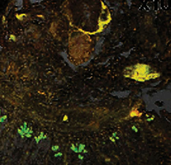
Image: Luminescent conjugated oligothiophene, h-FTAA staining of tissue showing amyloid deposits, yellow red fluorescence (Photo courtesy of Linköping University).
The amyloidoses comprise a large group of diseases characterized by extracellular deposits in various tissues and organs. These deposits, termed “amyloid,” result from misfolding and/or partial unfolding of proteins followed by their ordered aggregation into amyloid fibrils with specific β-sheet conformation.
Diagnosis of amyloidosis requires a tissue biopsy and the biopsy is assessed for evidence of characteristic amyloid deposits. The most useful stain in the diagnosis of amyloid is Congo red, which, combined with polarized light, makes the amyloid proteins appear apple-green on microscopy.
Scientists at the Linköping University (Sweden) and their colleagues identified patients diagnosed with amyloidosis in the amyloid collection of Uppsala Biobank (Sweden). They chose 53 paraffin-embedded tissue blocks with noticeable amyloid deposits from 50 patients. A second set of amyloid specimens was retrieved and included 61 paraffin-embedded tissue blocks with conspicuous amyloid deposits from 61 patients, including 24 women and 37 men. A control set of eight tissue samples were retrieved from five patients aged 69 to 78 years, comprising heart, liver and kidney. Tissues from a control group of 27, included kidney, stomach, heart, lung, ovary, pancreas and duodenum, which showed no interstitial amyloid deposits as assessed by Congo red staining and polarization microscopy.
Archived paraffin-embedded formalin fixed sections were processed and stained with a luminescent conjugated oligothiophene, h-FTAA, that stains rapidly and with high sensitivity and selectivity detects amyloid deposits in verified clinical samples from systemic amyloidosis patients with AA, AL and ATTR types; as well as in tissues laden with localized amyloidosis of AANF, AIAPP and ASem1 type. For the archived paraffin-embedded formalin fixed tissue specimens as well as dried fine needle aspired (FNA) abdominal fat biopsies it was found that a 1:5,000 staining solution corresponding to 0.2 mg/L of h-FTAA was optimal. Fluorescence images and spectra were recorded with a Leica DM6000 B fluorescence microscope (Leica Microsystems; Wetzlar, Germany).
The probe h-FTAA emitted yellow red fluorescence on binding to amyloid deposits, whereas no apparent staining was observed in surrounding tissue. The only functional structure stained with h-FTAA showing the amyloidotypic fluorescence spectrum was Paneth cell granules in intestine. Screening of 114 amyloid containing tissues derived from 107 verified (Congo red birefringence and/or immunohistochemistry) amyloidosis patients revealed complete correlation between h-FTAA and Congo red fluorescence. The majority of Congo red negative control cases (27 of 32, 85% specificity) were negative with h-FTAA. Small Congo red negative aggregates in kidney, liver, pancreas and duodenum were found by h-FTAA fluorescence in five control patients aged 72–83 years suffering from diverse diseases.
The authors concluded that conclude that h-FTAA is a fluorescent hypersensitive, rapid and powerful tool for identifying amyloid deposits in tissue sections. Use of h-FTAA can be exploited as a rapid complementary technique for accurate detection of amyloid in routine surgical pathology settings. The results also implicate the potential of the technique for detection of prodromal amyloidosis as well as for discovery of new amyloid-like protein aggregates in humans. The study was published on March 17, 2016, in Amyloid: The Journal of Protein Folding Disorders.
Related Links:
Linköping University
Leica Microsystems
Latest Pathology News
- AI Integrated With Optical Imaging Technology Enables Rapid Intraoperative Diagnosis
- HPV Self-Collection Solution Improves Access to Cervical Cancer Testing
- Hyperspectral Dark-Field Microscopy Enables Rapid and Accurate Identification of Cancerous Tissues
- AI Advancements Enable Leap into 3D Pathology
- New Blood Test Device Modeled on Leeches to Help Diagnose Malaria
- Robotic Blood Drawing Device to Revolutionize Sample Collection for Diagnostic Testing
- Use of DICOM Images for Pathology Diagnostics Marks Significant Step towards Standardization
- First of Its Kind Universal Tool to Revolutionize Sample Collection for Diagnostic Tests
- AI-Powered Digital Imaging System to Revolutionize Cancer Diagnosis
- New Mycobacterium Tuberculosis Panel to Support Real-Time Surveillance and Combat Antimicrobial Resistance
- New Method Offers Sustainable Approach to Universal Metabolic Cancer Diagnosis
- Spatial Tissue Analysis Identifies Patterns Associated With Ovarian Cancer Relapse
- Unique Hand-Warming Technology Supports High-Quality Fingertip Blood Sample Collection
- Image-Based AI Shows Promise for Parasite Detection in Digitized Stool Samples
- Deep Learning Powered AI Algorithms Improve Skin Cancer Diagnostic Accuracy
- Microfluidic Device for Cancer Detection Precisely Separates Tumor Entities
Channels
Clinical Chemistry
view channel
3D Printed Point-Of-Care Mass Spectrometer Outperforms State-Of-The-Art Models
Mass spectrometry is a precise technique for identifying the chemical components of a sample and has significant potential for monitoring chronic illness health states, such as measuring hormone levels... Read more.jpg)
POC Biomedical Test Spins Water Droplet Using Sound Waves for Cancer Detection
Exosomes, tiny cellular bioparticles carrying a specific set of proteins, lipids, and genetic materials, play a crucial role in cell communication and hold promise for non-invasive diagnostics.... Read more
Highly Reliable Cell-Based Assay Enables Accurate Diagnosis of Endocrine Diseases
The conventional methods for measuring free cortisol, the body's stress hormone, from blood or saliva are quite demanding and require sample processing. The most common method, therefore, involves collecting... Read moreMolecular Diagnostics
view channelBlood Proteins Could Warn of Cancer Seven Years before Diagnosis
Two studies have identified proteins in the blood that could potentially alert individuals to the presence of cancer more than seven years before the disease is clinically diagnosed. Researchers found... Read moreUltrasound-Aided Blood Testing Detects Cancer Biomarkers from Cells
Ultrasound imaging serves as a noninvasive method to locate and monitor cancerous tumors effectively. However, crucial details about the cancer, such as the specific types of cells and genetic mutations... Read moreHematology
view channel
Next Generation Instrument Screens for Hemoglobin Disorders in Newborns
Hemoglobinopathies, the most widespread inherited conditions globally, affect about 7% of the population as carriers, with 2.7% of newborns being born with these conditions. The spectrum of clinical manifestations... Read more
First 4-in-1 Nucleic Acid Test for Arbovirus Screening to Reduce Risk of Transfusion-Transmitted Infections
Arboviruses represent an emerging global health threat, exacerbated by climate change and increased international travel that is facilitating their spread across new regions. Chikungunya, dengue, West... Read more
POC Finger-Prick Blood Test Determines Risk of Neutropenic Sepsis in Patients Undergoing Chemotherapy
Neutropenia, a decrease in neutrophils (a type of white blood cell crucial for fighting infections), is a frequent side effect of certain cancer treatments. This condition elevates the risk of infections,... Read more
First Affordable and Rapid Test for Beta Thalassemia Demonstrates 99% Diagnostic Accuracy
Hemoglobin disorders rank as some of the most prevalent monogenic diseases globally. Among various hemoglobin disorders, beta thalassemia, a hereditary blood disorder, affects about 1.5% of the world's... Read moreImmunology
view channel.jpg)
AI Predicts Tumor-Killing Cells with High Accuracy
Cellular immunotherapy involves extracting immune cells from a patient's tumor, potentially enhancing their cancer-fighting capabilities through engineering, and then expanding and reintroducing them into the body.... Read more
Diagnostic Blood Test for Cellular Rejection after Organ Transplant Could Replace Surgical Biopsies
Transplanted organs constantly face the risk of being rejected by the recipient's immune system which differentiates self from non-self using T cells and B cells. T cells are commonly associated with acute... Read more
AI Tool Precisely Matches Cancer Drugs to Patients Using Information from Each Tumor Cell
Current strategies for matching cancer patients with specific treatments often depend on bulk sequencing of tumor DNA and RNA, which provides an average profile from all cells within a tumor sample.... Read more
Genetic Testing Combined With Personalized Drug Screening On Tumor Samples to Revolutionize Cancer Treatment
Cancer treatment typically adheres to a standard of care—established, statistically validated regimens that are effective for the majority of patients. However, the disease’s inherent variability means... Read moreMicrobiology
view channel
Integrated Solution Ushers New Era of Automated Tuberculosis Testing
Tuberculosis (TB) is responsible for 1.3 million deaths every year, positioning it as one of the top killers globally due to a single infectious agent. In 2022, around 10.6 million people were diagnosed... Read more
Automated Sepsis Test System Enables Rapid Diagnosis for Patients with Severe Bloodstream Infections
Sepsis affects up to 50 million people globally each year, with bacteraemia, formerly known as blood poisoning, being a major cause. In the United States alone, approximately two million individuals are... Read moreEnhanced Rapid Syndromic Molecular Diagnostic Solution Detects Broad Range of Infectious Diseases
GenMark Diagnostics (Carlsbad, CA, USA), a member of the Roche Group (Basel, Switzerland), has rebranded its ePlex® system as the cobas eplex system. This rebranding under the globally renowned cobas name... Read more
Clinical Decision Support Software a Game-Changer in Antimicrobial Resistance Battle
Antimicrobial resistance (AMR) is a serious global public health concern that claims millions of lives every year. It primarily results from the inappropriate and excessive use of antibiotics, which reduces... Read moreTechnology
view channel
New Diagnostic System Achieves PCR Testing Accuracy
While PCR tests are the gold standard of accuracy for virology testing, they come with limitations such as complexity, the need for skilled lab operators, and longer result times. They also require complex... Read more
DNA Biosensor Enables Early Diagnosis of Cervical Cancer
Molybdenum disulfide (MoS2), recognized for its potential to form two-dimensional nanosheets like graphene, is a material that's increasingly catching the eye of the scientific community.... Read more
Self-Heating Microfluidic Devices Can Detect Diseases in Tiny Blood or Fluid Samples
Microfluidics, which are miniature devices that control the flow of liquids and facilitate chemical reactions, play a key role in disease detection from small samples of blood or other fluids.... Read more
Breakthrough in Diagnostic Technology Could Make On-The-Spot Testing Widely Accessible
Home testing gained significant importance during the COVID-19 pandemic, yet the availability of rapid tests is limited, and most of them can only drive one liquid across the strip, leading to continued... Read moreIndustry
view channel
Danaher and Johns Hopkins University Collaborate to Improve Neurological Diagnosis
Unlike severe traumatic brain injury (TBI), mild TBI often does not show clear correlations with abnormalities detected through head computed tomography (CT) scans. Consequently, there is a pressing need... Read more
Beckman Coulter and MeMed Expand Host Immune Response Diagnostics Partnership
Beckman Coulter Diagnostics (Brea, CA, USA) and MeMed BV (Haifa, Israel) have expanded their host immune response diagnostics partnership. Beckman Coulter is now an authorized distributor of the MeMed... Read more_1.jpg)


_1.jpg)
.jpg)
