3D Imaging Method Determines Prostate Cancer Aggressiveness
|
By LabMedica International staff writers Posted on 23 Dec 2021 |

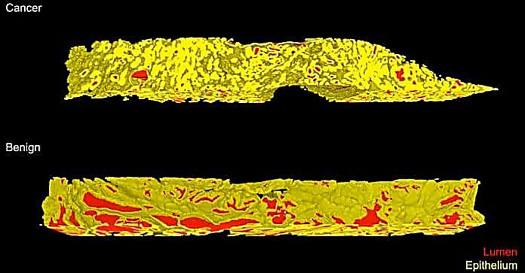
Image: A screenshot of a volume rendering of glands in two 3D biopsy samples from prostates (yellow: the outer walls of the gland; red: the fluid-filled space inside the gland). The cancer sample (top) shows smaller and more densely packed glands compared to the benign tissue sample (bottom) (Photo courtesy of Xie et al./Cancer Research)
Prostate cancer is the most common cancer for men and, for men in the USA, and it is the second leading cause of death. Prostate cancer treatment planning is largely dependent upon examination of core-needle biopsies. The microscopic architecture of the prostate glands forms the basis for prognostic grading by pathologists.
Some prostate cancers (PCas)might be slow-growing and can be monitored over time whereas others need to be treated right away. To determine how aggressive someone's cancer is, doctors look for abnormalities in histological specimens of biopsied tissue on a slide, but this 2D method makes it hard to properly diagnose borderline cases.
Biomedical Engineers at the University of Washington (Seattle, WA, USA) and their colleagues developed a workflow for non-destructive 3D pathology and computational analysis of whole prostate biopsies labeled with a rapid and inexpensive fluorescent analog of standard H&E staining. The team imaged 300 ex vivo biopsies extracted from 50 archived radical prostatectomy specimens, of which 118 biopsies contained cancer.
The biopsy cores were processed stained to mimic the typical staining used in the 2D method. The team then imaged each entire biopsy core using an open-top light-sheet microscope, which uses a sheet of light to optically “slice” through and image a tissue sample without destroying it. Multi-channel illumination was provided by a fourchannel digitally controlled laser package (Cobolt Skyra Lasers, HÜBNER Photonics, Kassel, Germany). Tissues were imaged at near Nyquist sampling of ∼0.44 μm/pixel. The volumetric imaging time was approximately 0.5 min per mm3 of tissue for each wavelength channel. This allowed each biopsy (~1 × 1 × 20 mm), stained with two fluorophores (T&E), to be imaged in ~20 minutes.
The scientists reported that the 3D images provided more information than a 2D image, specifically, details about the complex tree-like structure of the glands throughout the tissue. These additional features increased the likelihood that the computer would correctly predict a cancer's aggressiveness. They used new AI methods, including deep-learning image transformation techniques, to help manage and interpret the large datasets this project generated. The 3D glandular features in cancer biopsies were superior to corresponding 2D features for risk stratification of low- to intermediate-risk PCa patients based on their clinical biochemical recurrence (BCR) outcomes.
Jonathan Liu, PhD, a professor of mechanical engineering and of bioengineering and a senior author of the study, said, “We show for the first time that compared to traditional pathology, where a small fraction of each biopsy is examined in 2D on microscope slides, the ability to examine 100% of a biopsy in 3D is more informative and accurate. This is exciting because it is the first of hopefully many clinical studies that will demonstrate the value of non-destructive 3D pathology for clinical decision-making, such as determining which patients require aggressive treatments or which subsets of patients would respond best to certain drugs.”
The authors concluded that the results of this study support the use of computational 3D pathology for guiding the clinical management of prostate cancer. The study was published on December 1, 2021 in the journal Cancer Research.
Related Links:
University of Washington
HÜBNER Photonics
Latest Technology News
- Robotic Technology Unveiled for Automated Diagnostic Blood Draws
- ADLM Launches First-of-Its-Kind Data Science Program for Laboratory Medicine Professionals
- Aptamer Biosensor Technology to Transform Virus Detection
- AI Models Could Predict Pre-Eclampsia and Anemia Earlier Using Routine Blood Tests
- AI-Generated Sensors Open New Paths for Early Cancer Detection
- Pioneering Blood Test Detects Lung Cancer Using Infrared Imaging
- AI Predicts Colorectal Cancer Survival Using Clinical and Molecular Features
- Diagnostic Chip Monitors Chemotherapy Effectiveness for Brain Cancer
- Machine Learning Models Diagnose ALS Earlier Through Blood Biomarkers
- Artificial Intelligence Model Could Accelerate Rare Disease Diagnosis
Channels
Clinical Chemistry
view channel
New PSA-Based Prognostic Model Improves Prostate Cancer Risk Assessment
Prostate cancer is the second-leading cause of cancer death among American men, and about one in eight will be diagnosed in their lifetime. Screening relies on blood levels of prostate-specific antigen... Read more
Extracellular Vesicles Linked to Heart Failure Risk in CKD Patients
Chronic kidney disease (CKD) affects more than 1 in 7 Americans and is strongly associated with cardiovascular complications, which account for more than half of deaths among people with CKD.... Read moreMolecular Diagnostics
view channel
Diagnostic Device Predicts Treatment Response for Brain Tumors Via Blood Test
Glioblastoma is one of the deadliest forms of brain cancer, largely because doctors have no reliable way to determine whether treatments are working in real time. Assessing therapeutic response currently... Read more
Blood Test Detects Early-Stage Cancers by Measuring Epigenetic Instability
Early-stage cancers are notoriously difficult to detect because molecular changes are subtle and often missed by existing screening tools. Many liquid biopsies rely on measuring absolute DNA methylation... Read more
“Lab-On-A-Disc” Device Paves Way for More Automated Liquid Biopsies
Extracellular vesicles (EVs) are tiny particles released by cells into the bloodstream that carry molecular information about a cell’s condition, including whether it is cancerous. However, EVs are highly... Read more
Blood Test Identifies Inflammatory Breast Cancer Patients at Increased Risk of Brain Metastasis
Brain metastasis is a frequent and devastating complication in patients with inflammatory breast cancer, an aggressive subtype with limited treatment options. Despite its high incidence, the biological... Read moreHematology
view channel
New Guidelines Aim to Improve AL Amyloidosis Diagnosis
Light chain (AL) amyloidosis is a rare, life-threatening bone marrow disorder in which abnormal amyloid proteins accumulate in organs. Approximately 3,260 people in the United States are diagnosed... Read more
Fast and Easy Test Could Revolutionize Blood Transfusions
Blood transfusions are a cornerstone of modern medicine, yet red blood cells can deteriorate quietly while sitting in cold storage for weeks. Although blood units have a fixed expiration date, cells from... Read more
Automated Hemostasis System Helps Labs of All Sizes Optimize Workflow
High-volume hemostasis sections must sustain rapid turnaround while managing reruns and reflex testing. Manual tube handling and preanalytical checks can strain staff time and increase opportunities for error.... Read more
High-Sensitivity Blood Test Improves Assessment of Clotting Risk in Heart Disease Patients
Blood clotting is essential for preventing bleeding, but even small imbalances can lead to serious conditions such as thrombosis or dangerous hemorrhage. In cardiovascular disease, clinicians often struggle... Read moreImmunology
view channelBlood Test Identifies Lung Cancer Patients Who Can Benefit from Immunotherapy Drug
Small cell lung cancer (SCLC) is an aggressive disease with limited treatment options, and even newly approved immunotherapies do not benefit all patients. While immunotherapy can extend survival for some,... Read more
Whole-Genome Sequencing Approach Identifies Cancer Patients Benefitting From PARP-Inhibitor Treatment
Targeted cancer therapies such as PARP inhibitors can be highly effective, but only for patients whose tumors carry specific DNA repair defects. Identifying these patients accurately remains challenging,... Read more
Ultrasensitive Liquid Biopsy Demonstrates Efficacy in Predicting Immunotherapy Response
Immunotherapy has transformed cancer treatment, but only a small proportion of patients experience lasting benefit, with response rates often remaining between 10% and 20%. Clinicians currently lack reliable... Read moreMicrobiology
view channel
Comprehensive Review Identifies Gut Microbiome Signatures Associated With Alzheimer’s Disease
Alzheimer’s disease affects approximately 6.7 million people in the United States and nearly 50 million worldwide, yet early cognitive decline remains difficult to characterize. Increasing evidence suggests... Read moreAI-Powered Platform Enables Rapid Detection of Drug-Resistant C. Auris Pathogens
Infections caused by the pathogenic yeast Candida auris pose a significant threat to hospitalized patients, particularly those with weakened immune systems or those who have invasive medical devices.... Read moreTechnology
view channel
Robotic Technology Unveiled for Automated Diagnostic Blood Draws
Routine diagnostic blood collection is a high‑volume task that can strain staffing and introduce human‑dependent variability, with downstream implications for sample quality and patient experience.... Read more
ADLM Launches First-of-Its-Kind Data Science Program for Laboratory Medicine Professionals
Clinical laboratories generate billions of test results each year, creating a treasure trove of data with the potential to support more personalized testing, improve operational efficiency, and enhance patient care.... Read moreAptamer Biosensor Technology to Transform Virus Detection
Rapid and reliable virus detection is essential for controlling outbreaks, from seasonal influenza to global pandemics such as COVID-19. Conventional diagnostic methods, including cell culture, antigen... Read more
AI Models Could Predict Pre-Eclampsia and Anemia Earlier Using Routine Blood Tests
Pre-eclampsia and anemia are major contributors to maternal and child mortality worldwide, together accounting for more than half a million deaths each year and leaving millions with long-term health complications.... Read moreIndustry
view channelNew Collaboration Brings Automated Mass Spectrometry to Routine Laboratory Testing
Mass spectrometry is a powerful analytical technique that identifies and quantifies molecules based on their mass and electrical charge. Its high selectivity, sensitivity, and accuracy make it indispensable... Read more
AI-Powered Cervical Cancer Test Set for Major Rollout in Latin America
Noul Co., a Korean company specializing in AI-based blood and cancer diagnostics, announced it will supply its intelligence (AI)-based miLab CER cervical cancer diagnostic solution to Mexico under a multi‑year... Read more
Diasorin and Fisher Scientific Enter into US Distribution Agreement for Molecular POC Platform
Diasorin (Saluggia, Italy) has entered into an exclusive distribution agreement with Fisher Scientific, part of Thermo Fisher Scientific (Waltham, MA, USA), for the LIAISON NES molecular point-of-care... Read more


