Gel and Capillary Electrophoresis Compared for Human Proteins
|
By LabMedica International staff writers Posted on 12 May 2021 |


|
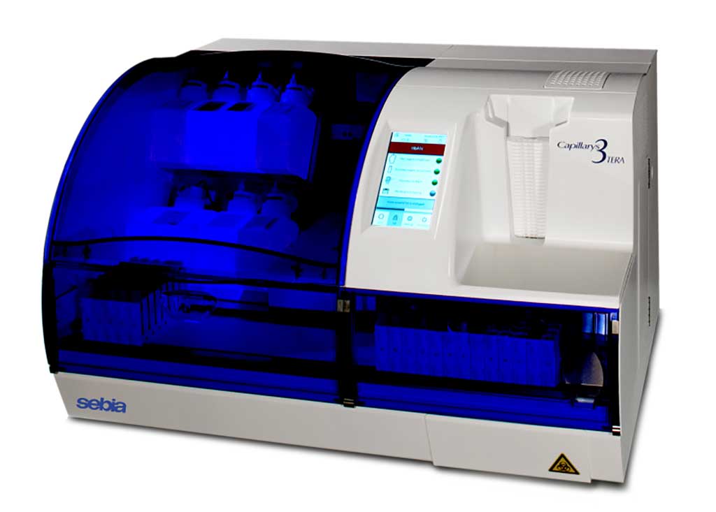
Image: The Capillarys 3 TERA is an automated analyzer based on capillary zone electrophoresis and UV detection for the quantitative analysis of Hba1c, proteins (serum and urine), immunotyping (serum and urine), and carbohydrate deficient transferrin (Photo courtesy of Sebia)
The electrophoresis of serum proteins remains a laboratory cornerstone for the diagnosis and monitoring of patients with plasma cell disorders, such as monoclonal gammopathy of undetermined significance, multiple myeloma, Waldenström macroglobulinemia, and AL amyloidosis.
Gel electrophoresis remains the method used by most clinical laboratories in the USA. However usage of gel electrophoresis is gradually decreasing while that of capillary electrophoresis is increasing. More recently, analysis of liquid media by capillary electrophoresis was developed and applied to the analysis of serum proteins for clinical purposes.
Laboratory Immunologists at The Johns Hopkins School of Medicine (Baltimore, MD, USA) tested all serum samples analyzed by the immunology laboratory over the course of eight days for routine clinical purposes during the months of October and November 2019, totaling 304 sera. There were 160 females (62 ± 16 years, range 19 -95) and 144 males (65 ± 14 years, range 23-97), with no significant difference in age according to sex.
Sera were first ran using the system currently in use, which consists of a thin-layer agarose gel, HYDRAGEL 30 Protein (E) (Sebia, Lisses, France) and the Sebia Hydrasys 2 semi-automated analyzer. Sera were then assayed on the Sebia Capillarys III TERA automated analyzer using CAPI 3 Protein (E) reagents. Serum immunofixation electrophoresis (SIFE) was performed in 214 of the total 304 sera because already ordered by the provider for most cases. Sera were pre-diluted according to the immunoglobulin G, A, and M concentrations, and then ran on the immunofixation system currently in use, which consists of a thin-layer agarose gel (HYDRAGEL IF 2/4, from Sebia) and the Hydrasys 2 semi-automated analyzer.
The scientists reported that Gel and capillary estimated the concentration of albumin, gamma region, and M-spikes nearly perfectly, and that of beta, alpha-2, and alpha-1 regions with excellent correlation. The two systems classified concordantly 268 of 304 sera (88% agreement) as having no, one, or two M-spikes, but differed in the remaining 36 sera (12%). Gel electrophoresis correctly identified M-spikes in 82 of 112 sera that were shown to have monoclonal band(s) by immunofixation (73% sensitivity), and correctly did not reveal M-spikes in 97/102 sera that had no immunofixation bands (95% specificity). Capillary achieved slightly higher sensitivity (85/112, 76%) and slightly lower specificity (94/102, 92%), but the two areas under the ROC curves were nearly identical at 0.84.
The authors concluded that Gel and capillary electrophoresis systems perform similarly to estimate the concentration of serum protein fractions and detect M-spikes. The Capillarys system has the disadvantages of a higher equipment cost and requiring a greater sample volume. On the other hand, Capillarys requires less involvement of laboratory technicians, offers a faster turn-around time, identifies the specimens they are being analyzed, does not depend upon the chemicals needed to stain and destain the gels, and is more amenable to remote reporting. The study was published on April 29, 2021 in the journal Practical Laboratory Medicine.
Related Links:
The Johns Hopkins School of Medicine
Sebia





Gel electrophoresis remains the method used by most clinical laboratories in the USA. However usage of gel electrophoresis is gradually decreasing while that of capillary electrophoresis is increasing. More recently, analysis of liquid media by capillary electrophoresis was developed and applied to the analysis of serum proteins for clinical purposes.
Laboratory Immunologists at The Johns Hopkins School of Medicine (Baltimore, MD, USA) tested all serum samples analyzed by the immunology laboratory over the course of eight days for routine clinical purposes during the months of October and November 2019, totaling 304 sera. There were 160 females (62 ± 16 years, range 19 -95) and 144 males (65 ± 14 years, range 23-97), with no significant difference in age according to sex.
Sera were first ran using the system currently in use, which consists of a thin-layer agarose gel, HYDRAGEL 30 Protein (E) (Sebia, Lisses, France) and the Sebia Hydrasys 2 semi-automated analyzer. Sera were then assayed on the Sebia Capillarys III TERA automated analyzer using CAPI 3 Protein (E) reagents. Serum immunofixation electrophoresis (SIFE) was performed in 214 of the total 304 sera because already ordered by the provider for most cases. Sera were pre-diluted according to the immunoglobulin G, A, and M concentrations, and then ran on the immunofixation system currently in use, which consists of a thin-layer agarose gel (HYDRAGEL IF 2/4, from Sebia) and the Hydrasys 2 semi-automated analyzer.
The scientists reported that Gel and capillary estimated the concentration of albumin, gamma region, and M-spikes nearly perfectly, and that of beta, alpha-2, and alpha-1 regions with excellent correlation. The two systems classified concordantly 268 of 304 sera (88% agreement) as having no, one, or two M-spikes, but differed in the remaining 36 sera (12%). Gel electrophoresis correctly identified M-spikes in 82 of 112 sera that were shown to have monoclonal band(s) by immunofixation (73% sensitivity), and correctly did not reveal M-spikes in 97/102 sera that had no immunofixation bands (95% specificity). Capillary achieved slightly higher sensitivity (85/112, 76%) and slightly lower specificity (94/102, 92%), but the two areas under the ROC curves were nearly identical at 0.84.
The authors concluded that Gel and capillary electrophoresis systems perform similarly to estimate the concentration of serum protein fractions and detect M-spikes. The Capillarys system has the disadvantages of a higher equipment cost and requiring a greater sample volume. On the other hand, Capillarys requires less involvement of laboratory technicians, offers a faster turn-around time, identifies the specimens they are being analyzed, does not depend upon the chemicals needed to stain and destain the gels, and is more amenable to remote reporting. The study was published on April 29, 2021 in the journal Practical Laboratory Medicine.
Related Links:
The Johns Hopkins School of Medicine
Sebia

Platinum Member
Automated Coagulation Analyzer
Hemolumi H6
Gold Member
NEW PRODUCT : SILICONE WASHING MACHINE TRAY COVER WITH VICOLAB SILICONE NET VICOLAB®
REGISTRED 682.9
New
Fully Automated Urinalysis System
DxU 1800 Fully Automated Urinalysis System
Urine Analyzer
respons® UDS100
Latest Technology News
- New Automation Workflow Streamlines NGS Library Preparation for High-Volume Labs
- ADLM Calls for CLIA Updates to Support Safe AI Use in Laboratory Medicine
- Multimodal AI Framework Aims to Guide Cancer Immunotherapy Decisions
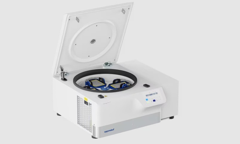
- New Multipurpose Centrifuge Combines High Capacity with Sustainable Cooling
- Bacterial Vesicle Expression System Streamlines Production of Cancer Diagnostic Proteins
- Autonomous Robotic System Receives FDA Authorization for Blood Collection
- Interoperable Data Platform Standardizes Multi-Cancer Blood Test Results
- Training Device Improves Accuracy of Pooled Molecular Diagnostics
- New CE-Certified Software Advances Whole-Genome Cancer Testing
- National Rare Disease Registry Standardizes Genetic and Clinical Data for Coordinated Care
- AI Platform Links Biomarker Results to Cancer Clinical Trials and Guidelines
- Agentic AI Platform Supports Genomic Decision-Making in Oncology
- Algorithm Panel Aids Liver Fibrosis Assessment and Liver Cancer Surveillance
- Mailed Screening Kits Help Reduce Colorectal Cancer Screening Gaps
- AI-Enabled Assistant Unifies Molecular Workflow Planning and Support
- AI Tool Automates Validation of Laboratory Software Configuration Changes
Channels
Clinical Chemistry
view channel
Breath Analysis Shows Promise for Distinguishing Cancerous from Benign Lung Nodules
Lung nodules are frequently detected during imaging, but distinguishing malignant from benign findings can require invasive procedures. Some patients undergo biopsies, bronchoscopy, or surgery before learning... Read more
Blood Biomarker Patterns Could Support Monitoring of Anti-Amyloid Treatment
Patients with early-stage Alzheimer’s disease may receive anti-amyloid therapy to clear amyloid protein deposits from the brain. These treatments can slow disease progression, but they do not cure the... Read more
Prehospital Blood Test Could Reduce Emergency Transfers for Chest Pain
Chest pain leads to ambulance transport to an emergency department for about 95% of patients, although only a minority have a serious cardiac condition. Troponin testing helps assess heart muscle damage,... Read more
First-of-Its-Kind Rapid Blood Test Gains CE Mark to Support Prehospital Stroke Triage
Rapid stroke identification before hospital arrival is critical because treatment efficacy declines with every minute of delay. However, definitive assessment typically requires hospital-based imaging,... Read moreMolecular Diagnostics
view channel
Long-Read Sequencing Test Identifies Genetic Causes in Inherited Muscle Disease
Inherited muscle diseases are highly heterogeneous and often progressive, leaving many patients without a molecular diagnosis despite extensive testing. Conventional assays typically target individual... Read more
Genomic Test Guides Extended Endocrine Therapy in Early Breast Cancer
Adjuvant endocrine therapy is standard for hormone receptor–positive early breast cancer, yet prolonged treatment brings side effects and adherence problems. Whether to continue beyond five years is a... Read more

Hematology
view channel
New Donor Genetic Marker May Help Predict Stem Cell Transplant Success
Donor selection for hematopoietic stem cell transplantation plays a major role in relapse risk and survival for patients with blood cancers and other blood disorders. Despite advances in genotyping, uncertainty... Read more
Updated Ferritin Thresholds Improve Detection of Iron Deficiency
Iron deficiency is one of the most common health conditions worldwide, yet its nonspecific symptoms can delay diagnosis for months. Variation in testing practices and ferritin thresholds may contribute... Read more
Microbiology
view channel
FDA Clears Rapid Phenotypic Antimicrobial Susceptibility System for Positive Blood Cultures
Bloodstream infections require prompt treatment, but antimicrobial susceptibility results often lag behind a positive blood culture. Conventional testing can take another 24 to 48 hours after a culture... Read more
New Urine Test Expands Mycotoxin Analysis to 31 Markers for Broader Exposure Assessment
Clinical evaluation of mold exposure increasingly relies on urinary mycotoxin testing, but limited marker coverage and metabolite masking can make results more difficult to interpret. Broader analysis... Read more
Pathology
view channel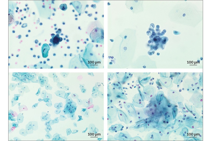
Simple Test Could Reduce the Need for Invasive Uterine Cancer Checks
Bleeding after menopause requires prompt investigation for uterine cancer, although most women who undergo testing do not have the disease. In the U.K., uterine cancer affects nearly 10,000 women each... Read more
Tumor Blood Vessel Features May Help Predict Colorectal Cancer Survival
Colorectal cancer outcomes vary widely, and tumor biology remains a key determinant of prognosis. Because neoplasms depend on a vascular supply, differences in intratumoral vessels may influence survival.... Read more
Multimodal AI Improves Breast Cancer Recurrence Risk Prediction Beyond Standard Genomic Testing
Predicting which patients with early-stage breast cancer will develop distant recurrence remains difficult, complicating decisions about long-term therapy and surveillance. Widely used genomic assays are... Read more
Technology
view channel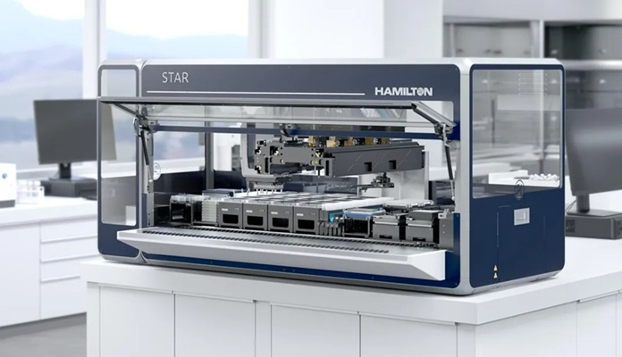
New Automation Workflow Streamlines NGS Library Preparation for High-Volume Labs
Demand for next-generation sequencing (NGS) continues to rise as laboratories process growing sample volumes, yet library preparation remains labor-intensive and a frequent source of variability.... Read more
ADLM Calls for CLIA Updates to Support Safe AI Use in Laboratory Medicine
Clinical laboratories increasingly use artificial intelligence to verify, interpret, and report results, but safeguards under the Clinical Laboratory Improvement Amendments (CLIA) were designed in 1992.... Read more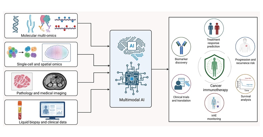
Industry
view channel
Collaboration Combines AI Cognitive Assessment and RNA Blood Testing for Earlier Alzheimer’s Detection
Alzheimer’s disease is often identified only after substantial neurodegeneration, partly because current diagnostic pathways are fragmented and difficult to scale. As treatment shifts toward earlier intervention,... Read more
Mayo Clinic Laboratories and Pathology Asia Expand Genomic Testing Across Asia-Pacific
Mayo Clinic Laboratories and Pathology Asia Holdings (PAH), together with subsidiary LifeStrands Genomics, announced a strategic investment and collaboration focused on expanding access to advanced diagnostics... Read more