Inexpensive LAMP-Based Schistosomiasis Tests Developed
|
By LabMedica International staff writers Posted on 12 Nov 2019 |


|
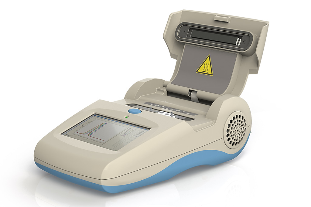
Image: The hand-held Genie III scanner has a two-color fluorescence excitation and detection system (Photo courtesy of OPTIGENE Ltd).
Schistosomiasis is one of the most prevalent Neglected Tropical Diseases, affecting approximately 250 million people worldwide. Schistosoma mansoni is the most important species causing human intestinal schistosomiasis.
Several polymerase chain reaction (PCR)-based molecular approaches, including conventional PCR (cPCR), real-time quantitative PCR (qPCR), droplet digital PCR (ddPCR), have been proven effective in detection of schistosome-derived DNA with more sensitivity than parasitological and serological methods, especially in chronic infections and in low transmission areas.
Scientists at the University of Salamanca (Salamanca, Spain) originally developed a loop-mediated isothermal amplification (LAMP) method for the detection of S. mansoni DNA (SmMIT-LAMP). They have now developed an important improvement for SmMIT-LAMP molecular assay, transforming it into a cold maintenance dry format suitable for potentially manufacturing as kit for ready-to-use for schistosomiasis diagnosis.
DNA from three patients’ tissue samples with microscopy-confirmed infection with S. mansoni was tested by LAMP including cutaneous and hepatic biopsies and an appendix biopsy. DNA was isolated from tissue samples using the QIAamp DNA Mini Kit (QIAGEN, Hilden, Germany). The real-time SmMIT-LAMP reactions were performed in 8-tube Genie Strips on a portable Genie III device (OPTIGENE Ltd., Horsham, UK) at 65 °C for 60 minutes followed up by 10 minutes at 80 °C. Amplicons were confirmed on 1.5% agarose gels when required.
The team reported that endpoint results at 65–70 minutes (counting 60 minutes for reaction plus 5–10 minutes of inactivation) were clearly observed with naked eye by adding the fluorescent dye SYBR Green I post-amplification. The positive LAMP reactions turned to green; otherwise, they remained orange. Correlation of positive colorimetric results with the typical ladder of multiple bands on agarose gels was clear.
Real-time SmMIT-LAMP was carried out on a portable device using the same reaction conditions, but testing including or not betaine in the master mix. Real-time reactions worked properly in both cases and a strong correlation between the signal of the fluorescent EvaGreen dye and electrophoresis was obtained. However, removing betaine from the mixture resulted on a 10 minute reduction in the amplification time while showing identical intensity of electrophoresis bands. Both fresh and desiccated SmMIT-LAMP mixtures yielded amplification signals for S. mansoni-positive control and tissue samples. Nevertheless, a delay in the appearance of positive results and a decrease in the fluorescence signals were observed when using desiccated mixtures.
The authors concluded that the one-step dry-up protocol is simpler and faster than those previously reported and allows maintaining the functionality for at least three weeks at RT and up to five months at 4 °C. Their work demonstrated an important improvement for SmMIT-LAMP molecular assay, transformed into a cold maintenance dry format suitable for manufacturing as kit for ready-to-use for S. mansoni DNA detection. The study was published on October 14, 2019 in the journal Scientific Reports.
Related Links:
University of Salamanca
QIAGEN
OPTIGENE Ltd




Several polymerase chain reaction (PCR)-based molecular approaches, including conventional PCR (cPCR), real-time quantitative PCR (qPCR), droplet digital PCR (ddPCR), have been proven effective in detection of schistosome-derived DNA with more sensitivity than parasitological and serological methods, especially in chronic infections and in low transmission areas.
Scientists at the University of Salamanca (Salamanca, Spain) originally developed a loop-mediated isothermal amplification (LAMP) method for the detection of S. mansoni DNA (SmMIT-LAMP). They have now developed an important improvement for SmMIT-LAMP molecular assay, transforming it into a cold maintenance dry format suitable for potentially manufacturing as kit for ready-to-use for schistosomiasis diagnosis.
DNA from three patients’ tissue samples with microscopy-confirmed infection with S. mansoni was tested by LAMP including cutaneous and hepatic biopsies and an appendix biopsy. DNA was isolated from tissue samples using the QIAamp DNA Mini Kit (QIAGEN, Hilden, Germany). The real-time SmMIT-LAMP reactions were performed in 8-tube Genie Strips on a portable Genie III device (OPTIGENE Ltd., Horsham, UK) at 65 °C for 60 minutes followed up by 10 minutes at 80 °C. Amplicons were confirmed on 1.5% agarose gels when required.
The team reported that endpoint results at 65–70 minutes (counting 60 minutes for reaction plus 5–10 minutes of inactivation) were clearly observed with naked eye by adding the fluorescent dye SYBR Green I post-amplification. The positive LAMP reactions turned to green; otherwise, they remained orange. Correlation of positive colorimetric results with the typical ladder of multiple bands on agarose gels was clear.
Real-time SmMIT-LAMP was carried out on a portable device using the same reaction conditions, but testing including or not betaine in the master mix. Real-time reactions worked properly in both cases and a strong correlation between the signal of the fluorescent EvaGreen dye and electrophoresis was obtained. However, removing betaine from the mixture resulted on a 10 minute reduction in the amplification time while showing identical intensity of electrophoresis bands. Both fresh and desiccated SmMIT-LAMP mixtures yielded amplification signals for S. mansoni-positive control and tissue samples. Nevertheless, a delay in the appearance of positive results and a decrease in the fluorescence signals were observed when using desiccated mixtures.
The authors concluded that the one-step dry-up protocol is simpler and faster than those previously reported and allows maintaining the functionality for at least three weeks at RT and up to five months at 4 °C. Their work demonstrated an important improvement for SmMIT-LAMP molecular assay, transformed into a cold maintenance dry format suitable for manufacturing as kit for ready-to-use for S. mansoni DNA detection. The study was published on October 14, 2019 in the journal Scientific Reports.
Related Links:
University of Salamanca
QIAGEN
OPTIGENE Ltd

Platinum Member
Automated Coagulation Analyzer
Hemolumi H6
Platinum Member
Integrated Biochemical & Immunological System
Biolumi CX Solution X10+C10
Gold Member
Nucleic Acid Extractor System
NEOS-96 XT
Latest Microbiology News
- Molecular Panel Gains WHO Emergency Use Listing to Support Ebola Outbreak Response
- Rapid Host-Response Test Aims to Predict Corticosteroid Benefit in Sepsis
- Oral and Lung Microbiome Changes Linked to Severe Long Covid Symptoms
- Study Finds Distinct Malaria Parasites Linked to Sickle Cell Trait Protection
- Rapid Urine Test Aids Diagnosis of Invasive Aspergillosis
- Nanodroplet CRISPR Technology Supports Rapid, Multiplexed Mycobacterial Identification
- Genomic Workflow Identifies Fungal Pathogens Before Blood Cultures Turn Positive
- FDA-Cleared Multiplex PCR Test Detects 13 Respiratory Pathogens in a Single Sample
- One-Hour Molecular Panel Expands Bloodstream Infection Testing for Gram-Negative Pathogens
- FDA Clears Rapid Phenotypic Antimicrobial Susceptibility System for Positive Blood Cultures
- New Urine Test Expands Mycotoxin Analysis to 31 Markers for Broader Exposure Assessment
- Collaboration Targets Blood-Based Ebola Detection Outside Central Laboratories
- Study Finds Frequent Overlap in Antibodies to Lyme and Other Tick-Borne Pathogens
- Early-Life Gut Microbiome Changes May Help Assess Type 1 Diabetes Risk
- FDA Breakthrough-Designated Urine Panel Aims to Shorten Pathogen and Susceptibility Testing
- Expanded Newborn Screening Program Increases Detection of Congenital Cytomegalovirus
Channels
Clinical Chemistry
view channel
Study Links Saliva-Based Epigenetic Signals to OCD Treatment Outcomes
Obsessive-compulsive disorder (OCD) causes unwanted, recurring thoughts and repetitive behaviors that can severely disrupt daily life. Exposure-based psychotherapy is among the most effective treatments,... Read more
New One-Step Syphilis Test Could Enable Earlier Detection and Faster Treatment
Syphilis can be difficult to diagnose because its symptoms may resemble other diseases and appear and disappear without treatment. Undetected infection can continue causing harm, spread to partners and... Read more
Hematology
view channel
New Myelofibrosis Guidelines Recommend Expanded Genetic Testing at Diagnosis
Myelofibrosis is a rare, life-threatening blood cancer affecting up to six per 100,000 people. The disease causes scarring in the bone marrow, limiting the production of healthy blood cells and potentially... Read more
Handheld Point-of-Care Test Aims to Enable Rapid Coagulation Assessment in Trauma
Severe blood loss and inadequate tissue oxygenation are leading causes of preventable death after traumatic injury. Blood products are essential in trauma care, but cold-chain storage, short shelf life,... Read more

Immunology
view channel
Immune Profiling Initiative Aims to Improve Bone Marrow Analysis in Leukemia
Acute myeloid leukemia (AML) is an aggressive blood cancer in which assessing disease burden after therapy can help guide clinical evaluation. Patients with measurable residual disease (MRD) after treatment... Read more
Immune Profiling Identifies Early Maternal Autoantibodies Linked to Preterm Birth Risk
A typical pregnancy lasts about 40 weeks, but labor begins prematurely in roughly one in 10 pregnancies. Preterm birth is the leading cause of newborn death worldwide, yet its underlying causes and which... Read more
Longitudinal Blood Testing Reveals Changes in Allergen Sensitization During Childhood
Food allergy is a major childhood health concern, and distinguishing persistent risk from transient sensitization can guide clinical decisions. Specific IgE antibodies may be detectable even when a child... Read more
Microbiology
view channel
Molecular Panel Gains WHO Emergency Use Listing to Support Ebola Outbreak Response
The ongoing Ebola Bundibugyo outbreak in the Democratic Republic of the Congo and neighboring countries has become one of the largest outbreaks of this Ebola virus species recorded to date.... Read more
Rapid Host-Response Test Aims to Predict Corticosteroid Benefit in Sepsis
Sepsis and severe infections are difficult to treat because patients with similar clinical presentations may have very different underlying immune responses. Corticosteroids are commonly used in severe... Read more
Pathology
view channel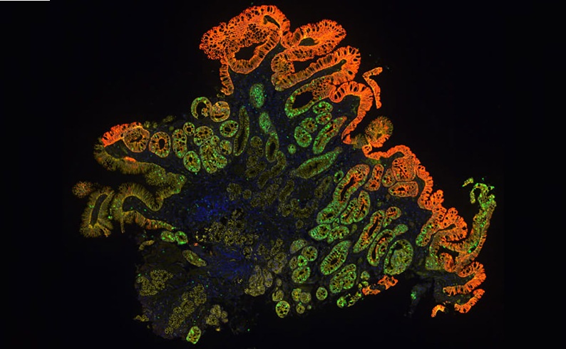
Molecular Testing Refines Barrett’s Esophagus Progression Risk Beyond Biopsy Findings
Barrett’s esophagus is a precancerous condition associated with increased risk of esophageal adenocarcinoma. Routine endoscopic biopsy findings guide surveillance and therapy, but histopathology may not... Read more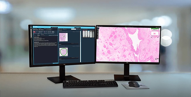
Updated Digital Pathology Platform Expands AI Integration for Cancer Diagnosis
Digital pathology is reshaping how laboratories manage case review, image analysis, and collaboration. As diagnostic workflows become more complex, pathology teams need platforms that can support digital... Read more
Technology
view channel
Laser-Based Swab Analysis Shows Promise for Detecting Disease-Linked Odor Patterns
Disease-related changes in volatile organic compounds can alter body odor, producing measurable patterns in exhaled breath and bodily fluids. Current analytical methods can be complex, time-consuming,... Read more
Laser-Enhanced Assay Boosts Sensitivity for Colorectal Cancer Biomarker Detection
Colorectal cancer is the third most commonly diagnosed cancer and the second leading cause of cancer-related death worldwide. Early detection remains critical, but cancer biomarkers can produce only faint... Read more

Industry
view channel
Hologic to Broaden Breast Cancer Testing Portfolio Through Reveal Genomics Acquisition
Hologic (Marlborough, MA, USA) has signed a definitive agreement to acquire Reveal Genomics (Barcelona, Spain), a precision oncology company developing genomic diagnostic tests to help personalize cancer treatment.... Read more