Immunoassay Detects Severe Fever with Thrombocytopenia Syndrome Virus
|
By LabMedica International staff writers Posted on 25 Apr 2016 |
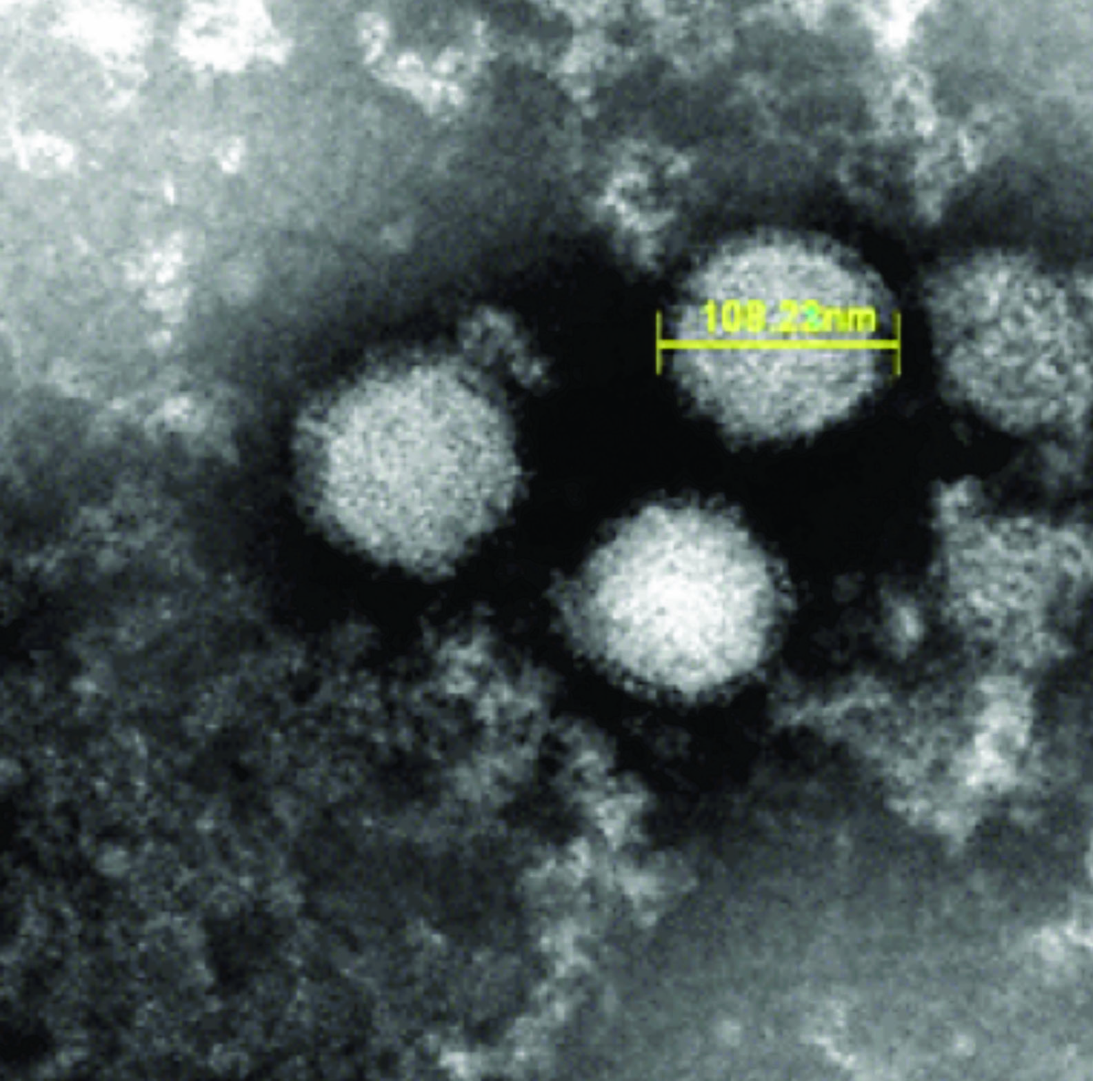
Image: A scanning electron micrograph (SEM) of severe fever with thrombocytopenia syndrome virus (Photo courtesy of the Japanese National Institute of Infectious Diseases).
Severe fever with thrombocytopenia syndrome (SFTS) is a tick-borne infectious disease with a high case fatality rate, and is caused by the SFTS virus (SFTSV) and the disease is endemic to China, South Korea, and Japan.
The viral ribonucleic acid (RNA) level in sera of patients with SFTS is known to be strongly associated with outcomes and therefore virological SFTS diagnosis with high sensitivity and specificity are required in disease endemic areas.
Scientists at the Japanese National Institute of Infectious Diseases (Tokyo, Japan) and their colleagues collected 63 serum samples from 55 acute phase patients suspected of SFTS in Japan. Viral gene detection by the quantitative reverse transcription polymerase chain reaction (qRT-PCR) and viral antibody detection by immunoglobulin G (IgG) enzyme-linked immunosorbent assay (ELISA) and/or indirect fluorescent antibody (IFA) were conducted to diagnose SFTS. From 55 patients, 34 of these were diagnosed as having SFTSV. Serum samples obtained from 18 healthy donors were used to establish the cut-off value of the IgG ELISA. Serum samples used for IgG ELISA were inactivated under the UV light in the biosafety cabinet for one hour.
The investigators generated novel monoclonal antibodies (MAbs) against the SFTSV nucleocapsid (N) protein and developed a sandwich antigen (Ag)-capture enzyme-linked immunosorbent assay (ELISA) for the detection of N protein of SFTSV using MAb and polyclonal antibody as capture and detection antibodies, respectively. The Ag-capture ELISAs were read using an optical density at 405 nm (OD405) was measured against a reference of 490 nm using a Model 680 Microplate Reader (Bio-Rad Laboratories Inc.; Hercules, CA, USA). The Ag-capture system was capable of detecting at least 350 to 1,220 50% Tissue Culture Infective Dose (TCID50)/100 μL/well from the culture supernatants of various SFTSV strains.
All 24 serum samples (100%) containing high copy numbers of viral RNA more than 105 copies/mL) showed a positive reaction in the Ag-capture ELISA, whereas 12 out of 15 serum samples (80%) containing low copy numbers of viral RNA (less than 105 copies/mL) showed a negative reaction in the Ag-capture ELISA. Among these Ag-capture ELISA- negative 12 samples, nine (75%) were positive for IgG antibodies against SFTSV. The authors conclude that the newly developed Ag-capture ELISA is useful for SFTS diagnosis in acute phase patients with high levels of viremia. The study was published on April 5, 2016, in the journal Public Library of Science Neglected Tropical Diseases.
Related Links:
Japanese National Institute of Infectious Diseases
Bio-Rad Laboratories
Latest Microbiology News
- Three-Test Panel Launched for Detection of Liver Fluke Infections
- Rapid Test Promises Faster Answers for Drug-Resistant Infections
- CRISPR-Based Technology Neutralizes Antibiotic-Resistant Bacteria
- Comprehensive Review Identifies Gut Microbiome Signatures Associated With Alzheimer’s Disease
- AI-Powered Platform Enables Rapid Detection of Drug-Resistant C. Auris Pathogens
- New Test Measures How Effectively Antibiotics Kill Bacteria
- New Antimicrobial Stewardship Standards for TB Care to Optimize Diagnostics
- New UTI Diagnosis Method Delivers Antibiotic Resistance Results 24 Hours Earlier
- Breakthroughs in Microbial Analysis to Enhance Disease Prediction
- Blood-Based Diagnostic Method Could Identify Pediatric LRTIs
- Rapid Diagnostic Test Matches Gold Standard for Sepsis Detection
- Rapid POC Tuberculosis Test Provides Results Within 15 Minutes
- Rapid Assay Identifies Bloodstream Infection Pathogens Directly from Patient Samples
- Blood-Based Molecular Signatures to Enable Rapid EPTB Diagnosis
- 15-Minute Blood Test Diagnoses Life-Threatening Infections in Children
- High-Throughput Enteric Panels Detect Multiple GI Bacterial Infections from Single Stool Swab Sample
Channels
Clinical Chemistry
view channel
Electronic Nose Smells Early Signs of Ovarian Cancer in Blood
Ovarian cancer is often diagnosed at a late stage because its symptoms are vague and resemble those of more common conditions. Unlike breast cancer, there is currently no reliable screening method, and... Read more
Simple Blood Test Offers New Path to Alzheimer’s Assessment in Primary Care
Timely evaluation of cognitive symptoms in primary care is often limited by restricted access to specialized diagnostics and invasive confirmatory procedures. Clinicians need accessible tools to determine... Read moreMolecular Diagnostics
view channel
New Blood Test Predicts Who Will Most Likely Live Longer
As people age, it becomes increasingly difficult to determine who is likely to maintain stable health and who may face serious decline. Traditional indicators such as age, cholesterol, and physical activity... Read more
Genetic Test Predicts Radiation Therapy Risk for Prostate Cancer Patients
External beam radiation therapy is widely used to treat localized prostate cancer, which has a five-year survival rate exceeding 99%. However, more than 20% of patients develop persistent urinary side... Read moreHematology
view channel
Rapid Cartridge-Based Test Aims to Expand Access to Hemoglobin Disorder Diagnosis
Sickle cell disease and beta thalassemia are hemoglobin disorders that often require referral to specialized laboratories for definitive diagnosis, delaying results for patients and clinicians.... Read more
New Guidelines Aim to Improve AL Amyloidosis Diagnosis
Light chain (AL) amyloidosis is a rare, life-threatening bone marrow disorder in which abnormal amyloid proteins accumulate in organs. Approximately 3,260 people in the United States are diagnosed... Read moreMicrobiology
view channel
Three-Test Panel Launched for Detection of Liver Fluke Infections
Parasitic liver fluke infections remain endemic in parts of Asia, where transmission commonly occurs through consumption of raw freshwater fish or aquatic plants. Chronic infection is a well-established... Read more
Rapid Test Promises Faster Answers for Drug-Resistant Infections
Drug-resistant pathogens continue to pose a growing threat in healthcare facilities, where delayed detection can impede outbreak control and increase mortality. Candida auris is notoriously difficult to... Read more
CRISPR-Based Technology Neutralizes Antibiotic-Resistant Bacteria
Antibiotic resistance has accelerated into a global health crisis, with projections estimating more than 10 million deaths per year by 2050 as drug-resistant “superbugs” continue to spread.... Read more
Comprehensive Review Identifies Gut Microbiome Signatures Associated With Alzheimer’s Disease
Alzheimer’s disease affects approximately 6.7 million people in the United States and nearly 50 million worldwide, yet early cognitive decline remains difficult to characterize. Increasing evidence suggests... Read morePathology
view channel
Urine Specimen Collection System Improves Diagnostic Accuracy and Efficiency
Urine testing is a critical, non-invasive diagnostic tool used to detect conditions such as pregnancy, urinary tract infections, metabolic disorders, cancer, and kidney disease. However, contaminated or... Read more
AI-Powered 3D Scanning System Speeds Cancer Screening
Cytology remains a cornerstone of cancer detection, requiring specialists to examine bodily fluids and cells under a microscope. This labor-intensive process involves inspecting up to one million cells... Read moreTechnology
view channel
Blood Test “Clocks” Predict Start of Alzheimer’s Symptoms
More than 7 million Americans live with Alzheimer’s disease, and related health and long-term care costs are projected to reach nearly USD 400 billion in 2025. The disease has no cure, and symptoms often... Read more
AI-Powered Biomarker Predicts Liver Cancer Risk
Liver cancer, or hepatocellular carcinoma, causes more than 800,000 deaths worldwide each year and often goes undetected until late stages. Even after treatment, recurrence rates reach 70% to 80%, contributing... Read more
Robotic Technology Unveiled for Automated Diagnostic Blood Draws
Routine diagnostic blood collection is a high‑volume task that can strain staffing and introduce human‑dependent variability, with downstream implications for sample quality and patient experience.... Read more
ADLM Launches First-of-Its-Kind Data Science Program for Laboratory Medicine Professionals
Clinical laboratories generate billions of test results each year, creating a treasure trove of data with the potential to support more personalized testing, improve operational efficiency, and enhance patient care.... Read moreIndustry
view channel
QuidelOrtho Collaborates with Lifotronic to Expand Global Immunoassay Portfolio
QuidelOrtho (San Diego, CA, USA) has entered a long-term strategic supply agreement with Lifotronic Technology (Shenzhen, China) to expand its global immunoassay portfolio and accelerate customer access... Read more


