New Microfluidics Method to Speed Up Blood Analyses
Posted on 21 Aug 2024
Researchers have developed a new method to accelerate and potentially scale up the process of separating particles in fluids, a technique that could prove useful for analyzing cancer cells from blood.
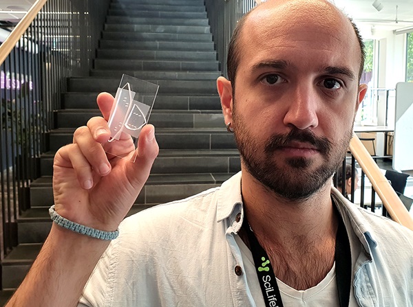
This speedier and more precise method of elasto-inertial microfluidics was developed by a team led by researchers at KTH Royal Institute of Technology (Stockholm, Sweden) and involves controlling the movement of tiny particles in fluids by leveraging both the fluid's elastic properties and the inertial forces arising from fluid movement. The microfluidic device features specially engineered channels that accommodate larger volumes of fluid rapidly, making it ideal for applications requiring quick, continuous particle separation. These channels efficiently sort and line up particles, essential for distinguishing different particle types.

This high precision is achieved through the use of specially formulated fluids with high polymer concentrations, giving the fluid viscoelastic properties similar to egg whites that can both flow and rebound. This combination of forces allows for precise control over particle movement. The study, published in Nature Microsystems & Nanoengineering, found that larger particles are more manageable and maintain focus even as fluid flow increases. Smaller particles, however, require optimal flow rates to stay in line, though control can improve under appropriate conditions. The improved technique offers a diverse range of potential uses in medical testing and can help quickly sort cells or other particles in blood samples.
“We showed how the sample throughput can be increased within our microfluidic channel,” said Selim Tanriverdi, a PhD student at KTH and lead author of the study. “This would lower the process time for blood analysis, which is crucial for a patient.”
Related Links:
KTH Royal Institute of Technology


 Analyzer.jpg)


