AI Application in Pathology Reveals Novel Insights in Endometrial Cancer Diagnostics
Posted on 19 Dec 2022
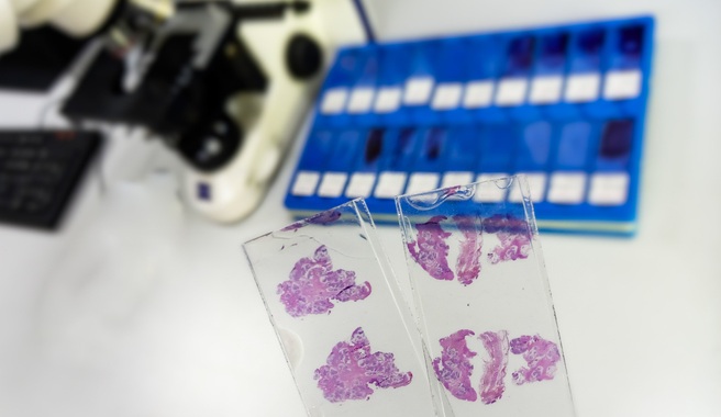
Endometrial carcinoma is the most common cancer of the gynecologic tract. Now, researchers have shown the power of artificial intelligence (AI) can be applied to endometrial carcinoma microscopy images, offering novel insights that could improve diagnosis and treatment of uterine cancer.
In the past years, researchers at Leiden University (Leiden, the Netherlands) had played a leading role in the development of a novel tumor classification system based on molecular alterations, resulting in four endometrial cancer subtypes. This time, the team set out to investigate if it was possible to predict these molecular classes, based on microscopy-images alone. The researchers applied artificial intelligence on microscopy images of thousands of endometrial carcinoma images from patients that participated in the study.
")
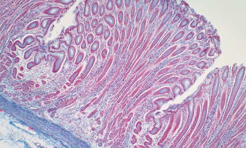
The team developed a model that robustly predicts the four molecular classes of endometrial carcinomas based on one (hematoxylin and eosin)-stained microscopy slide image, which is the standard histological stain used in diagnostics for assessment of tumor grading and histological subtyping. This model was not “a black-box”, but through reverse-engineering the researchers were able to show which image-features were relevant for its predictions. The model provided the team with important novel insights that can be utilized in future studies to further improve diagnostics, prognostication, and management of endometrial cancer patients.
“The application of AI in pathology is emerging,” said Dr. Tjalling Bosse at Leiden University. “In this project we studied the morphology of tumors that shared the same molecular alteration to better understand the effect these changes have on the appearance of the tumor. With this work, the computer model has directed us to areas in- and outside the tumor that are important.”
“In cancer diagnostics, the number of variables (molecular, tumor morphology, patient data) has increased exponentially and has complexified patient prognosis prediction,” added Sarah Fremond. “Through training unbiased AI models, AI predictions can also teach pathologists in return by, for instance, identifying novel morphological details on microscopy slide images with prognostic value.”
Related Links:
Leiden University