COVID-19 Lung Damage Caused by Persistence of Abnormal Cells
By LabMedica International staff writers
Posted on 19 Nov 2020
Several uncertainties still relate to the involvement of other organs in COVID-19. Besides indirect multi-organ injury, a few reports have suggested the possibility of direct injury caused by viral replication in brain, heart and kidney.Posted on 19 Nov 2020
Over the last couple of months a few studies have explored the lung pathology caused by COVID-19 infection in a small number of patients. The pattern that has emerged is that COVID-19 lung disease causes diffuse alveolar damage (DAD) which is also present in other conditions of acute respiratory distress syndrome (ARDS), including SARS.

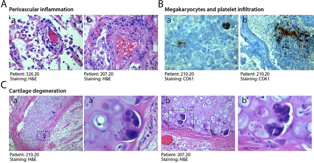
Image: Histological features of COVID-19 lungs (Photo courtesy of the University of Trieste).
A team of Medical Scientists at the University of Trieste (Trieste, Italy) analyzed the organs of 41 patients who died of COVID-19 at the University Hospital of Trieste. The team took lung, heart, liver, and kidney samples to examine the behavior of the virus. Of the 41 patients, six required intensive care, while 35 were hospitalized in either other hospital units or local nurseries until death. The average age of patients was 77 and 84 years for males and females, respectively. Hypertension, chronic cardiac disease, dementia, diabetes and cancer were the most common comorbidities. All patients eventually died of clinical acute respiratory distress syndrome.
At pathological examination, the investigators found that all cases exhibited lung damage and the lungs appeared congested at macroscopic examination. Meanwhile, histological analysis of all cases revealed gross destruction of the normal lung architecture, consistent with a condition of diffuse alveolar damage with edema, intra-alveolar fibrin deposition with hyaline membranes and hemorrhage. This was accompanied by occlusion of alveolar spaces due to cell delamination. Loss of cellular integrity was also confirmed by the presence of karyorrhexis, indicative of ongoing cellular death.
Further, in situ RNA hybridization for the detection of the SARS-CoV-2 genome indicated that the alterations in the lung were concomitant with persistent viral infection of pneumocytes and endothelial cells. RNA-positive pneumocytes were found to be largely present in the lungs of 10 of 11 tested individuals. The team said presence of abundant cytoplasmic RNA signals and expression of the Spike protein in the lungs after 30-40 days from diagnosis in the study suggests ongoing replication and postulates a continuous pathogenetic role of viral infection.
The scientists also noted that also noted the presence of anomalous epithelial cells among the cases which were characterized by abnormally large cytoplasm and, very commonly, by the presence of bi- of multi-nucleation. Presence of these dysmorphic cells was detected in 87% of patients. They added that the dysmorphic cells very often showed features of syncytia, characterized by several nuclei with an ample cytoplasm surrounded by a single plasma membrane. Most of these syncytia-forming, dysmorphic cells were bona fide pneumocytes.
The authors concluded that COVID-19 is a unique disease characterized by extensive lung thrombosis, long-term persistence of viral RNA in pneumocytes and endothelial cells, along with the presence of infected cell syncytia. Several of COVID-19 features might be consequent to the persistence of virus-infected cells for the duration of the disease. The study was published on November 3, 2020 in the journal EBioMedicine.
Related Links:
University of Trieste


