Non-Invasive Imaging Detects Cancer at Molecular Level
By LabMedica International staff writers
Posted on 20 Aug 2019
For cancer patients the presence of metastases dictates the staging assessment, which in turn defines the appropriate treatment path selected. For gynecological malignancies, like ovarian carcinoma, it is of immense importance to differentiate between localized and metastatic disease status as that drastically affects management.Posted on 20 Aug 2019
For in situ, real time diagnosis, novel imaging modalities that offer metabolic and structural information at the cellular and subcellular level can be of great help, especially since these modalities are being progressively incorporated in probes and micro-endoscopes that allow intra-vital access to organs that lie deeper in the body.
.")
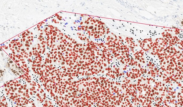
Image: Scientists combined multiphoton microscopy with automated image and statistical analysis algorithms to distinguish between healthy and diseased tissue. In this image, collected in a completely label-free, noninvasive manner, collagen is colored green while ovarian metastatic cell clusters are presented in red (Photo courtesy of Tufts University).
Biomedical scientists at Tufts University (Medford, MA, USA) and their colleagues collected samples from eight patients who underwent open laparotomy as part of routine medical care. Post completion of all intra-abdominal procedures of the operation, eight biopsies of healthy parietal peritoneum and if present of four peritoneal metastases were collected from each patient. All lesions were evaluated by a pathologist using standard hematoxylin and eosin histology.
The tissues were imaged employing a multiphoton laser scanning microscope to generate intrinsic fluorescence and second harmonic generation (SHG) images at 755 nm and 900 nm excitation respectively with signal emission collected at 460 ± 20 and 525 ± 25 nm. Laser light was focused on the sample using a 25x objective (0.9 NA / water-immersion), and neutral density filters were employed to achieve a power of 25–35 mW. At least two to three random fields per tissue were evaluated, reaching a total of 30 and 11 images for the healthy and metastatic biopsy tissue groups, respectively (512 × 512 pixels; 600-micron field of view; resolution of 1.17 microns per pixel). Imaging was focused within a depth of ∼20-100 microns from the mesothelial surface of the tissues.
The team found that healthy tissues displayed large variations in contrast and correlation features as a function of distance, corresponding to repetitive, increased local intensity fluctuations. Metastatic tissue images exhibited decreased contrast and correlation related values, representing more uniform intensity patterns and smaller fibers, indicating the destruction of the healthy stroma by the cancerous infiltration. Analyzing 41 images acquired from the biopsies, the technique correctly classified 40 out of 41 images (an accuracy of 97.5%). A total of 11 samples were correctly classified as metastatic (100% sensitivity) and 29 of 30 were correctly classified as healthy (96.6% specificity).
Dimitra Pouli, MD, PhD, a Pathology Resident and co-author of the study, said, “The method utilized in this work identifies in a completely label-free manner cellular and tissue features at the microscopic level, essentially acting like a biopsy without a knife,” The study was published in the August 2019 issue of the journal Biomedical Optics Express.
Related Links:
Tufts University
 Assay.jpg)
Gold Member
H-FABP Assay
Heart-Type Fatty Acid-Binding Protein Assay


All-in-One Molecular System
AIO M160

Prefilled Tubes
Prefilled 5.0ml Tubes