Noninvasive AI-Enabled Raman Probe Distinguishes Skin Cancers from Normal Tissue
Posted on 05 Oct 2026
Skin cancer is the most common cancer in the United States and among the most common worldwide. Nearly 1.5 million new cases were diagnosed globally in 2024, including nearly 340,000 melanomas. Many malignant, benign, and precancerous lesions can appear similar, while biopsy remains invasive and costly. New findings demonstrate a noninvasive optical approach that could help assess suspicious lesions.
Researchers at Florida Atlantic University (FAU; Boca Raton, FL, USA) are evaluating Raman spectroscopy-based optical diagnostics of skin cancer combined with machine learning. The approach uses Raman spectroscopy to generate a molecular fingerprint by analyzing how light scatters when it interacts with molecules in tissue. This provides information about chemical composition without removing or specially preparing tissue.
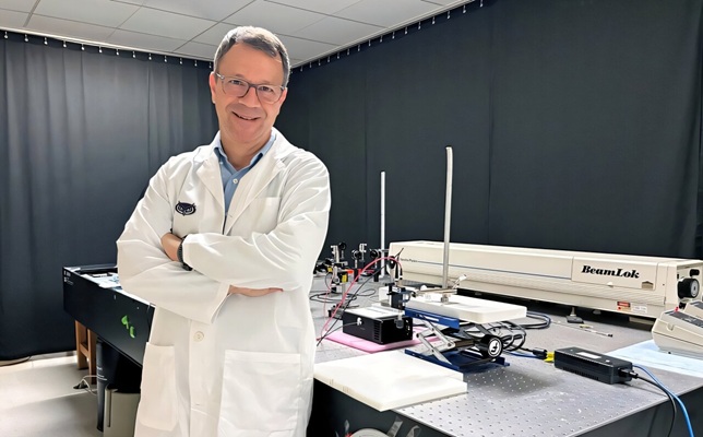
The team tested a mobile Raman spectroscopy system equipped with a 785-nanometer diode laser and handheld probe. The ex vivo analysis included more than 50 clinical samples, comprising basal cell carcinoma, squamous cell carcinoma, and normal skin. Researchers generated nearly 1,000 Raman spectra and evaluated multiple machine-learning methods to determine how well spectral data could distinguish among the three tissue types.
Several machine-learning approaches identified patterns in Raman spectra that separated normal skin from cancerous tissue. K-nearest neighbors and support vector machine classifiers achieved the highest overall test accuracy, at about 84%. The support vector machine reached 78.7% sensitivity and 88.6% specificity, while a shallow neural network achieved 80.8% accuracy and the highest receiver operating characteristic area under the curve, or ROC AUC, at 0.910.
The analysis showed that normal tissue could be distinguished relatively well from basal cell carcinoma and squamous cell carcinoma, although the two cancer types had greater overlap in their molecular signatures. Cancerous samples tended to show stronger protein-related signals, while normal tissue showed stronger lipid-related signals. The study was published in Proceedings of SPIE as part of Advanced Chemical Microscopy for Life Science and Translational Medicine 2026.
“Our results are preliminary, but they point toward a future in which a rapid, noninvasive measurement could help guide clinical decisions and potentially reduce unnecessary biopsies,” said Andrew Terentis, Ph.D., professor and chair of the Department of Chemistry and Biochemistry at Florida Atlantic University.
Related Links
Florida Atlantic University