New Microscope Promises to Speed Up Medical Diagnostics
Posted on 19 Sep 2025
Traditional microscopes are designed for flat samples, yet real-life specimens, such as tissue slides, are often curved or uneven. This mismatch forces researchers to rely on scanning methods or costly optics, slowing imaging and limiting resolution. Now, a new approach enables extremely large, high-resolution images of curved samples in a single snapshot, offering faster insights for biology applications.
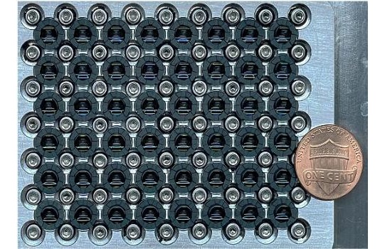
Researchers at Duke University (Durham, NC, USA) have introduced a new microscope called PANORAMA, which captures gigapixel-scale images without mechanical scanning. The device combines a telecentric lens originally designed for chip manufacturing with a large tube lens that projects the sample image onto an array of 48 small cameras. Each camera covers a portion of the sample and can be independently focused, ensuring clarity across uneven surfaces.

The system, detailed in Optics Letters, creates submicron detail — as fine as 1/120 the diameter of a human hair — over areas comparable to the size of a U.S. dime. In demonstrations, the device imaged a prepared rat brain tissue slice in one snapshot, producing a 630 MP image with neurons and dendrites clearly visible. Tests on onion skin placed over a curved surface also showed crisp brightfield and fluorescence images, with stained nuclei and cell walls sharply resolved.
By eliminating slow focus stacking and tedious tile scanning, the new microscope dramatically increases throughput and flexibility. It could be applied in pathology to scan biopsy slides at cellular resolution almost instantly. Future improvements aim to expand its field of view to capture entire petri dishes, add automated focusing, and enable 3D reconstructions or live video imaging.
“This tool can be used wherever large-area, detailed imaging is needed,” said Haitao Chen, a doctoral student at Duke University. “For instance, in medical pathology, it could scan entire tissue slides, such as those from a biopsy, at cellular resolution almost instantly.”
Related Links:
Duke University


