Combined Maternal and Fetal Gene Variants Predict Pre-Eclampsia Risk
By LabMedica International staff writers
Posted on 26 Mar 2020
A genetic signature that combines certain maternal and fetal gene variants shows promise as a means for assessing risk of pregnant women to develop pre-eclampsia.Posted on 26 Mar 2020
Pre-eclampsia (PE) is a disorder of pregnancy characterized by the onset of high blood pressure and often a significant amount of protein in the urine. When it arises, the condition begins after 20 weeks of pregnancy. In severe disease there may be red blood cell breakdown, a low blood platelet count, impaired liver function, kidney dysfunction, swelling, shortness of breath due to fluid in the lungs, or visual disturbances. Pre-eclampsia increases the risk of poor outcomes for both the mother and the baby. If left untreated, it may result in seizures at which point it is known as eclampsia. Ten to 15% of maternal mortality is associated with pre-eclampsia and eclampsia.

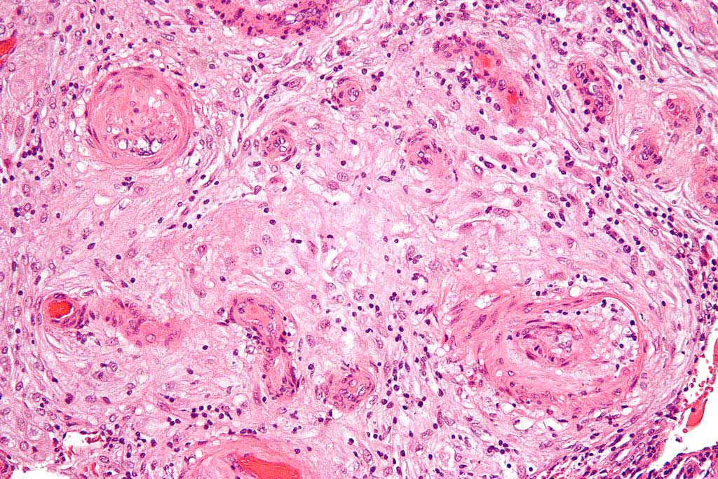
Image: High magnification micrograph of hypertrophic decidual vasculopathy, as seen in pregnancy-induced hypertension (Photo courtesy of Wikimedia Commons)
Systemic complement activation levels are increased during pregnancy compared to non-pregnant women of childbearing age. In PE, systemic complement levels are further increased, and higher complement deposition has been observed on placentas. In this light, investigators at Baylor Medical College (Houston, TX, USA) hypothesized that combinations of common SNPs in maternal and fetal complement genes predisposed women to PE.
To test this theory, the investigators screened a retrospective cohort of PE pregnancies to identify SNPs (single-nucleotide polymorphisms) in one fetal and two maternal complement genes and compared their frequencies with those from normal pregnancies. Since C3 is central effector molecule in the complement cascade, and CD46 and factor H regulate C3 activation, they selected CD46 (fetal) and C3, factor H (maternal) for sequencing in this study.
The investigators reported that they had identified a total of nine common SNPs. Minor allele frequencies of two fetal CD46 SNPs were significantly higher in PE. Furthermore, fetal CD46 variants and maternal CFH/C3 variants were highly prevalent in PE patients compared to normal pregnancies. Placental complement deposition and maternal alternative pathway 50 (AP50) values were higher in PE pregnancies. Irrespective of disease status, two CD46 variants were associated with reduced placental CD46 expression and one CFH variant was associated with increased maternal AP50 values.
The investigators concluded with the hope that this PE genetic signature could be used in the future to identify women at risk.
The study was published in the March 16, 2020, online edition of the journal Scientific Reports.
Related Links:
Baylor College of Medicine


