30-Minute 3D Histology Tool Supports Intraoperative Glioma Margin Assessment
Posted on 19 Aug 2026
Glioma surgery depends on distinguishing infiltrative tumor from functional brain tissue, but microscopic spread beyond visible margins makes this difficult. Conventional frozen sections provide rapid guidance but sample only a few two-dimensional planes and can be affected by artifacts and sampling bias. Three-dimensional histology preserves tissue architecture but typically requires hours to days, limiting intraoperative use. New findings demonstrate a 30-minute workflow that generates deep 3D histology to support margin assessment during glioma surgery.
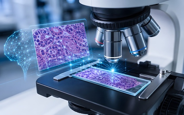
Researchers at Fudan University have developed ULTRA (ultrarapid cleared stimulated Raman with AI), a platform that integrates rapid tissue clearing, stimulated Raman scattering (SRS) microscopy, and artificial intelligence to visualize surgical specimens volumetrically without conventional staining or sectioning. The workflow compresses deep 3D histological analysis into roughly 30 minutes. It is designed to present tissue morphology in a hematoxylin and eosin (H&E)-like format that pathologists can readily interpret.
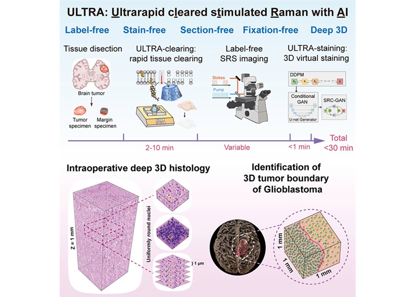
ULTRA begins by clearing fresh or fixed brain tissue for millimeter-scale volumetric imaging. Label-free stimulated Raman scattering (SRS) microscopy then captures intrinsic vibrational signals from chemical bonds, while AI modules reconstruct and virtually stain the data to generate H&E-like 3D histology. The AI pipeline includes depth-aware denoising to restore signals at increasing imaging depths, a conditional generative adversarial network that infers a protein channel from the lipid channel to reduce acquisition burden, and a virtual H&E staining model that renders the resulting images in an interpretable format.
In a study of surgical glioma specimens from 17 patients, including astrocytoma, oligodendroglioma, glioblastoma, childhood diffuse hemispheric glioma, and low-grade glioma, the platform revealed continuous 3D pathological features such as nuclear atypia, microcystic change, abnormal mitoses, microvascular proliferation, and necrosis. Mouse brain and other organ tissues were also used to validate tissue clearing, imaging depth, signal restoration, and preservation of tissue architecture. For intraoperative margin assessment, approximately 1 mm³ of glioblastoma tissue could be processed in about 30 minutes.
An ULTRAscore derived from cellularity analysis and a 3D convolutional neural network segmented volumes into dense tumor, sparse infiltrative tumor, and nontumor regions, highlighting depth-dependent heterogeneity at margins. Independent pathologists reviewing single 2D sections from the same volumes missed tumor present in adjacent planes, with miss rates of 20% and 23%. In benchmarking, a 3D convolutional neural network outperformed a 2D model, with an area under the curve (AUC) of 0.965 versus 0.909.
The study also showed detection of tumor in radiographically ambiguous edema regions and in areas appearing normal on navigation and negative on neurophysiological monitoring, suggesting specimen-based complementation of intraoperative imaging.
The work, conducted by teams from Fudan University, Huashan Hospital of Fudan University, Zhongshan Hospital of Fudan University, Beijing Neurosurgical Institute, Capital Medical University, and collaborators, is published in Cell on August 3, 2026. The authors emphasize methodological limits, including axial point-spread-function anisotropy, computational speed of diffusion-based models, potential tissue-specific optimization, and the current concentration of SRS systems in specialized centers, and note that large prospective trials are needed before clinical outcomes claims or replacement of frozen section can be made.
“Conventional intraoperative pathology often relies on a limited number of two-dimensional sections, while tumors themselves are highly heterogeneous three-dimensional entities. The goal of ULTRA is to obtain a more complete 3D histological view within a time frame close to intraoperative pathology, without destroying the tissue. We hope this technology will help doctors better understand glioma infiltration margins in the future and provide more tissue-level information for intraoperative decision-making,” said Dr. Lixue Shi of Fudan University.
Related Links
Fudan University