Digital Pathology Tool Predicts Breast Cancer Outcomes and Therapy Response
Posted on 22 Jul 2026
Breast cancer prognosis often depends on microscopic assessment of tumor architecture, a process that can be subjective and lead to variable predictions across patient groups. Quantitative measures that capture tissue organization could help laboratories deliver more consistent risk estimates and guide therapy selection. Many gene and protein markers also show fluctuating performance across racial and ethnic groups, underscoring the need for robust alternatives. A new study shows that topology-based measurements of tumor structure can improve prediction of outcomes and therapy response.
Columbia University Irving Medical Center researchers and collaborators developed a set of topology-based biomarkers that convert visual patterns in breast tumor tissue into continuous numerical scores. The approach applies mathematical tools from topology to formalize the “order versus disorder” that pathologists have long evaluated by eye. Investigators report that these scores transform qualitative impressions of tissue organization into reproducible, quantitative readouts.
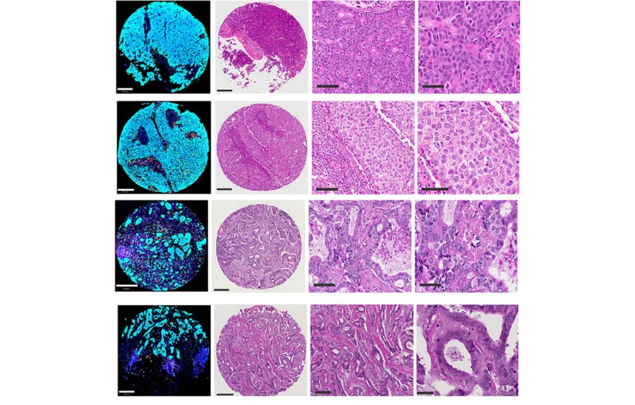
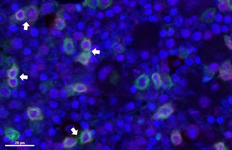
The method uses persistent homology to analyze digital images, focusing on how tumor and immune cells are spatially arranged rather than solely on which cells are present. Using multiplex immunofluorescence, the team mapped coordinates for tumor cells, immune cells, and programmed death-ligand 1 (PD-L1) expression. Computational models then quantified architectural patterns across the tissue, yielding topology-based scores that capture organization on a continuous scale.
Study data included samples from more than 550 breast cancer patients in a racially diverse North Carolina cohort. Higher topology scores, reflecting more organized tissue structure, were associated with longer survival and more favorable outcomes. The topology-based biomarkers outperformed several conventional approaches and remained highly predictive in both non-Hispanic Black and non-Hispanic white patient groups.
Investigators also integrated the topology measurements with gene expression to generate topology-derived gene signatures that predicted therapy response in independent breast cancer clinical trial datasets. Analyses linked low topology scores to biological pathways involved in metabolism, immune suppression, and epithelial-to-mesenchymal transition, with an observed connection to the metabolic enzyme IL4I1 and aryl hydrocarbon receptor signaling. The findings are published in Cancer Research, and the authors note that additional validation and clinical studies will be needed before routine clinical use.
Researchers are working to extend the approach from multiplex immunofluorescence to standard pathology slides used widely in clinical practice. If successful, the method could complement existing workflows by bringing quantitative measures of tissue architecture into digital pathology analysis. The team highlights the potential for broader access to advanced diagnostics if these computational tools can be applied to commonly available slide preparations.
"For more than a century, pathology has relied on recognizing patterns visually. What's exciting now is that we can begin to mathematically define those patterns and use them predictively. The long-term potential is that advanced cancer diagnostics could become faster, more quantitative, more accurate and far more broadly accessible than they are today," said Kevin Gardner, M.D., Ph.D., chair of the department of pathology and cell biology at Columbia University Irving Medical Center and senior author of the study.
Related Links
Columbia University Irving Medical Center