Microfluidic Device Captures Tumor-Specific Extracellular Vesicles
By LabMedica International staff writers
Posted on 21 Mar 2018
A microfluidic device has been developed that can capture glioblastoma-derived extracellular vesicles, with high specificity, using very small blood samples, which would be useful for pediatric patients.Posted on 21 Mar 2018
The microfluidic channels in the device contain a cocktail of antibodies that are specific for molecules found on glioblastoma-derived extracellular vesicles, meaning the vesicles are captured as they pass through the channels.
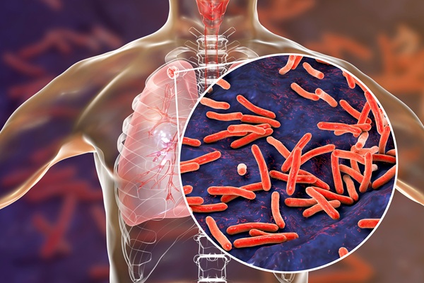
 released from a patient’s tumor and captured on the surfaces of the EVHB-Chip (Photo courtesy of Professor Shannon Stott, PhD).")
Image: Extracellular vesicles (red) released from a patient’s tumor and captured on the surfaces of the EVHB-Chip (Photo courtesy of Professor Shannon Stott, PhD).
Scientists at Massachusetts General Hospital (Charlestown, MA, USA) and their colleagues collected blood samples from a total of 13 brain cancer patients and six healthy donors were included in this study. Microfluidic devices consisted of 8-Channel herringbone structures were fabricated using standard photolithography and different strategies were tested for optimal configuration of capture antibodies on the surface of the microfluid device.
Isolated extracellular vesicles (EVs) were quantified using a tunable resistive pulse sensing (TRPS) qNano instrument. EV’s were isolated with immobilized with streptavidin-coated magnetic particles. The cyclic olefin copolymer (COC) The COC microfluidic device allowed direct imaging of captured EVs. Micrographs were captured with an LSM510 confocal microscope equipped with a ×63 Zeiss Plan-APOCHROMAT oil objective. RNA was isolated and quantified. Digital polymerase chain reaction, library preparation for RNA sequencing and RNA sequencing analysis was also performed.
The team reported that the sensitive analytical microfluidic platform (EVHB-Chip) that enables tumor-specific EV-RNA isolation within three hours. Using the EVHB-Chip, They achieved 94% tumor-EV specificity, a limit of detection of 100 EVs per μL, and a 10-fold increase in tumor RNA enrichment in comparison to other methods. This approach allowed for the subsequent release of captured tumor EVs, enabling downstream characterization and functional studies. Processing serum and plasma samples from glioblastoma multiforme (GBM) patients, they detected the mutant Type III epidermal growth factor receptor (EGFRvIII) messenger RNA (mRNA).
Shannon L. Stott, PhD, an Assistant Professor of Medicine and the lead investigator of the study, said, “Our device’s ability to sort tumor-specific extracellular vesicles out from the billions of extracellular vesicles carried through the blood stream may lead to the development of much-needed diagnostic and monitoring tools for this and other hard-to-treat cancers.” The study was published online on January 12, 2018, in the journal Nature Communications.
Related Links:
Massachusetts General Hospital

Gold Member
Aspiration System
VACUSAFE


Pipette Calibration System
Artel PCS®

Japanese Encephalitis Test
Japanese Encephalitis Virus Real Time PCR Kit