AI Tool to Transform Skin Cancer Detection with Near-Perfect Accuracy
Posted on 26 Nov 2025
Melanoma continues to be one of the most difficult skin cancers to diagnose because it often resembles harmless moles or benign lesions. Traditional AI tools depend heavily on dermoscopic images alone, overlooking important clinical details such as a patient’s age, sex, or lesion location, all of which can influence diagnostic accuracy. These limitations underscore the need for more integrated diagnostic approaches. Now, a new multimodal AI solution allows combining image features with patient context for more accurate detection of melanoma.
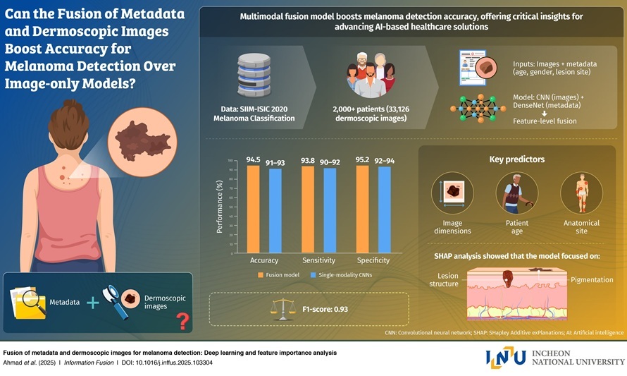
Researchers at Incheon National University (Incheon, South Korea), in collaboration with the University of the West of England (Bristol, UK) and other leading institutions, have created a deep learning model that combines dermoscopic imaging with clinical metadata to better distinguish melanoma from benign lesions. Their system was trained using the SIIM-ISIC dataset, which includes more than 33,000 paired dermoscopic images and patient information.

The model integrates visual features with details such as patient age, lesion size, and anatomical site to capture patterns that image-only systems may miss. This multimodal fusion allows the network to identify subtle diagnostic cues and align them with patient-specific risk factors. Using the large-scale dataset, the model achieved a diagnostic accuracy of 94.5% and an F1-score of 0.94, outperforming commonly used image-based architectures like ResNet-50 and EfficientNet.
Feature importance analysis showed that incorporating patient metadata meaningfully strengthened predictions and improved model transparency. The study highlighted that factors such as lesion location and patient age contributed significantly to melanoma classification. These findings, published in Information Fusion, demonstrate the potential of multimodal systems to reduce misclassification and improve reliability compared to image-only AI tools.
The combination of visual and clinical features provides a more comprehensive picture of melanoma risk and supports more reliable diagnostic output. The multimodal approach could be integrated into clinical workflows to support dermatologists in early melanoma identification, especially in settings where specialist access is limited. Its structure also makes it suitable for deployment in smartphone-based screening apps and telemedicine platforms. The technology offers a way to broaden early detection efforts and reduce reliance solely on visual assessment.
Future development may adapt the model into real-world diagnostic systems that analyze both images and basic patient inputs to guide early intervention. By linking machine intelligence with clinical context, this approach can help create more personalized, accurate, and accessible skin cancer diagnostics.
“The model is not merely designed for academic purposes. It could be used as a practical tool that could transform real-world melanoma screening,” said Prof. Gwangill Jeon. “This research can be directly applied to developing an AI system that analyzes both skin lesion images and basic patient information to enable early detection of melanoma.”
Related Links:
Incheon National University
University of the West of England


