Novel Surrogate Marker Described for Pancreatic Neuroendocrine Tumors
|
By LabMedica International staff writers Posted on 19 May 2022 |

Tumors of the endocrine pancreas are a collection of tumor cell types collectively referred to as PanNETs. These tumors originate in islet cells. Although they may be similar or identical in histologic appearance to carcinoid tumors of the gastrointestinal tract, differences in their underlying biology and likely differences in response to therapeutic agents suggest that they should be treated and investigated as a distinct entity.
Pancreatic neuroendocrine tumor (PanNET) is the second most common solid neoplasm of the pancreas, accounting for 3% to 4% of all pancreas neoplasms. The incidence of this tumor has increased owing to advances in various imaging techniques and increasing awareness. Surgical resection is the main treatment option for localized PanNETs, both functioning and nonfunctioning, without distant metastasis.
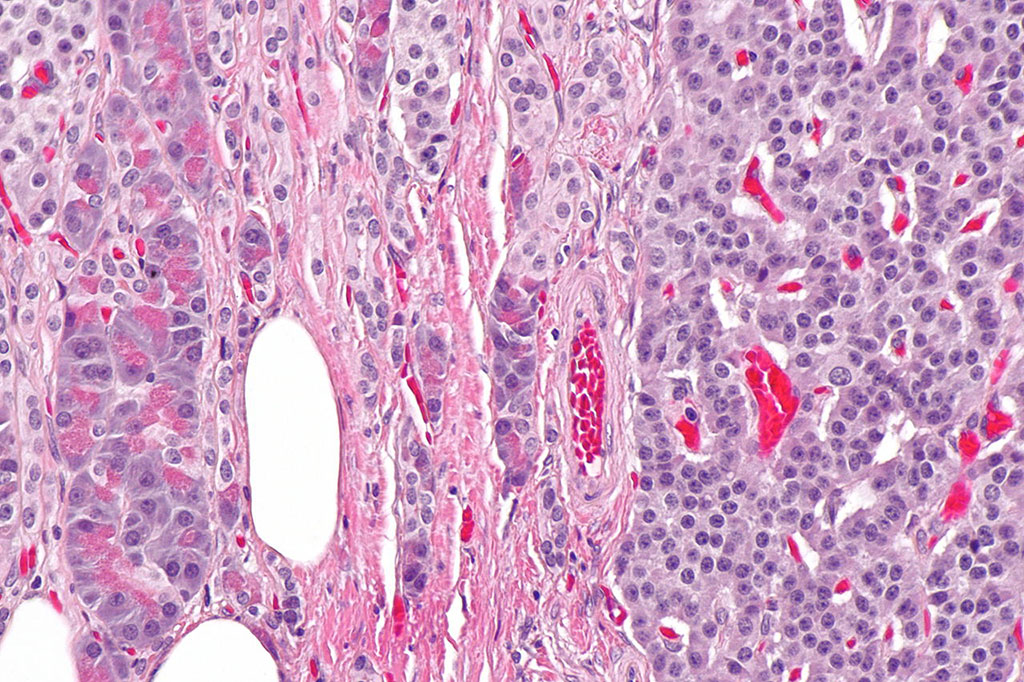
Pathologists at the University of Ulsan College of Medicine (Seoul, Republic of Korea) assessed the significance of tumor border, a well-known prognostic indicator in other cancers, in the PanNETs. They evaluated the macroscopic growth pattern (expansile [Exp] versus infiltrative [Inf]) and the microscopic tumor border (pushing [Pus] versus Inf) of 203 surgically resected PanNETs and compared them with other clinicopathologic factors.
Growth pattern and tumor border were evaluated at the junctions between tumor and normal pancreatic parenchyma or between tumor and other adjacent organs by both gross and microscopic examination. First, macroscopic growth pattern was evaluated in PanNET cases with available gross images of the cut surface of the PanNETs and was classified as expansile or infiltrative pattern. Expansile growth pattern was defined when the tumor had well-delineated margin from adjacent parenchyma. Microscopic tumor border was categorized as pushing or infiltrative. When the tumor boundary was well circumscribed under microscopic examination, this was referred to as a pushing border. Tissue microarray (TMA) was constructed from archived, formalin-fixed, paraffin-embedded tissue blocks with a manual tissue microarrayer (UNITMA Co Ltd, Seoul, Korea).
The scientists reported that based on macroscopic growth pattern, 83 cases had Exp patterns whereas 84 had Inf patterns. According to microscopic tumor border, 122 PanNETs had Pus borders whereas 81 had Inf borders. Combining macroscopic growth pattern and microscopic tumor border, 65 PanNETs had Exp/Pus, 34 had Inf/Pus, 18 had Exp/Inf, and 50 had Inf/Inf status. PanNETs with Inf/Inf status were associated with higher tumor grade, pT classification, and American Joint Committee on Cancer stage grouping; lymph node metastasis; and lymphovascular and perineural invasion.. Patients with PanNET having Inf/Inf status had significantly shorter overall survival (OS) and recurrence-free survival (RFS). Further, using multivariate analysis, Inf/Inf status was identified as an independent poor prognostic factor of OS and RFS.
The authors concluded that the combined Inf/Inf status was observed in approximately 25% of PanNETs and was associated with aggressive biological behavior and short OS and RFS. Therefore, assessing combined macroscopic growth pattern and microscopic tumor border can provide additional information regarding survival and recurrence in PanNET patients. The study was published on May 9, 2022 in the journal Archives of Pathology and Laboratory Medicine.
Related Links:
University of Ulsan College of Medicine
UNITMA Co Ltd
Latest Pathology News
- Robotic Blood Drawing Device to Revolutionize Sample Collection for Diagnostic Testing
- Use of DICOM Images for Pathology Diagnostics Marks Significant Step towards Standardization
- First of Its Kind Universal Tool to Revolutionize Sample Collection for Diagnostic Tests
- AI-Powered Digital Imaging System to Revolutionize Cancer Diagnosis
- New Mycobacterium Tuberculosis Panel to Support Real-Time Surveillance and Combat Antimicrobial Resistance
- New Method Offers Sustainable Approach to Universal Metabolic Cancer Diagnosis
- Spatial Tissue Analysis Identifies Patterns Associated With Ovarian Cancer Relapse
- Unique Hand-Warming Technology Supports High-Quality Fingertip Blood Sample Collection
- Image-Based AI Shows Promise for Parasite Detection in Digitized Stool Samples
- Deep Learning Powered AI Algorithms Improve Skin Cancer Diagnostic Accuracy
- Microfluidic Device for Cancer Detection Precisely Separates Tumor Entities
- Virtual Skin Biopsy Determines Presence of Cancerous Cells
- AI Detects Viable Tumor Cells for Accurate Bone Cancer Prognoses Post Chemotherapy
- First Ever Technique Identifies Single Cancer Cells in Blood for Targeted Treatments
- Innovative Blood Collection Device Overcomes Common Obstacles Related to Phlebotomy
- Intra-Operative POC Device Distinguishes Between Benign and Malignant Ovarian Cysts within 15 Minutes
Channels
Clinical Chemistry
view channel
3D Printed Point-Of-Care Mass Spectrometer Outperforms State-Of-The-Art Models
Mass spectrometry is a precise technique for identifying the chemical components of a sample and has significant potential for monitoring chronic illness health states, such as measuring hormone levels... Read more.jpg)
POC Biomedical Test Spins Water Droplet Using Sound Waves for Cancer Detection
Exosomes, tiny cellular bioparticles carrying a specific set of proteins, lipids, and genetic materials, play a crucial role in cell communication and hold promise for non-invasive diagnostics.... Read more
Highly Reliable Cell-Based Assay Enables Accurate Diagnosis of Endocrine Diseases
The conventional methods for measuring free cortisol, the body's stress hormone, from blood or saliva are quite demanding and require sample processing. The most common method, therefore, involves collecting... Read moreMolecular Diagnostics
view channel
Urine Test to Revolutionize Lyme Disease Testing
Lyme disease is the most common animal-to-human transmitted disease in the United States, with around 476,000 people diagnosed and treated annually, and its incidence has been increasing.... Read more
Simple Blood Test Could Enable First Quantitative Assessments for Future Cerebrovascular Disease
Cerebral small vessel disease is a common cause of stroke and cognitive decline, particularly in the elderly. Presently, assessing the risk for cerebral vascular diseases involves using a mix of diagnostic... Read more
New Genetic Testing Procedure Combined With Ultrasound Detects High Cardiovascular Risk
A key interest area in cardiovascular research today is the impact of clonal hematopoiesis on cardiovascular diseases. Clonal hematopoiesis results from mutations in hematopoietic stem cells and may lead... Read moreHematology
view channel
Next Generation Instrument Screens for Hemoglobin Disorders in Newborns
Hemoglobinopathies, the most widespread inherited conditions globally, affect about 7% of the population as carriers, with 2.7% of newborns being born with these conditions. The spectrum of clinical manifestations... Read more
First 4-in-1 Nucleic Acid Test for Arbovirus Screening to Reduce Risk of Transfusion-Transmitted Infections
Arboviruses represent an emerging global health threat, exacerbated by climate change and increased international travel that is facilitating their spread across new regions. Chikungunya, dengue, West... Read more
POC Finger-Prick Blood Test Determines Risk of Neutropenic Sepsis in Patients Undergoing Chemotherapy
Neutropenia, a decrease in neutrophils (a type of white blood cell crucial for fighting infections), is a frequent side effect of certain cancer treatments. This condition elevates the risk of infections,... Read more
First Affordable and Rapid Test for Beta Thalassemia Demonstrates 99% Diagnostic Accuracy
Hemoglobin disorders rank as some of the most prevalent monogenic diseases globally. Among various hemoglobin disorders, beta thalassemia, a hereditary blood disorder, affects about 1.5% of the world's... Read moreImmunology
view channel
Diagnostic Blood Test for Cellular Rejection after Organ Transplant Could Replace Surgical Biopsies
Transplanted organs constantly face the risk of being rejected by the recipient's immune system which differentiates self from non-self using T cells and B cells. T cells are commonly associated with acute... Read more
AI Tool Precisely Matches Cancer Drugs to Patients Using Information from Each Tumor Cell
Current strategies for matching cancer patients with specific treatments often depend on bulk sequencing of tumor DNA and RNA, which provides an average profile from all cells within a tumor sample.... Read more
Genetic Testing Combined With Personalized Drug Screening On Tumor Samples to Revolutionize Cancer Treatment
Cancer treatment typically adheres to a standard of care—established, statistically validated regimens that are effective for the majority of patients. However, the disease’s inherent variability means... Read moreMicrobiology
view channelEnhanced Rapid Syndromic Molecular Diagnostic Solution Detects Broad Range of Infectious Diseases
GenMark Diagnostics (Carlsbad, CA, USA), a member of the Roche Group (Basel, Switzerland), has rebranded its ePlex® system as the cobas eplex system. This rebranding under the globally renowned cobas name... Read more
Clinical Decision Support Software a Game-Changer in Antimicrobial Resistance Battle
Antimicrobial resistance (AMR) is a serious global public health concern that claims millions of lives every year. It primarily results from the inappropriate and excessive use of antibiotics, which reduces... Read more
New CE-Marked Hepatitis Assays to Help Diagnose Infections Earlier
According to the World Health Organization (WHO), an estimated 354 million individuals globally are afflicted with chronic hepatitis B or C. These viruses are the leading causes of liver cirrhosis, liver... Read more
1 Hour, Direct-From-Blood Multiplex PCR Test Identifies 95% of Sepsis-Causing Pathogens
Sepsis contributes to one in every three hospital deaths in the US, and globally, septic shock carries a mortality rate of 30-40%. Diagnosing sepsis early is challenging due to its non-specific symptoms... Read moreTechnology
view channel
New Diagnostic System Achieves PCR Testing Accuracy
While PCR tests are the gold standard of accuracy for virology testing, they come with limitations such as complexity, the need for skilled lab operators, and longer result times. They also require complex... Read more
DNA Biosensor Enables Early Diagnosis of Cervical Cancer
Molybdenum disulfide (MoS2), recognized for its potential to form two-dimensional nanosheets like graphene, is a material that's increasingly catching the eye of the scientific community.... Read more
Self-Heating Microfluidic Devices Can Detect Diseases in Tiny Blood or Fluid Samples
Microfluidics, which are miniature devices that control the flow of liquids and facilitate chemical reactions, play a key role in disease detection from small samples of blood or other fluids.... Read more
Breakthrough in Diagnostic Technology Could Make On-The-Spot Testing Widely Accessible
Home testing gained significant importance during the COVID-19 pandemic, yet the availability of rapid tests is limited, and most of them can only drive one liquid across the strip, leading to continued... Read moreIndustry
view channel_1.jpg)
Thermo Fisher and Bio-Techne Enter Into Strategic Distribution Agreement for Europe
Thermo Fisher Scientific (Waltham, MA USA) has entered into a strategic distribution agreement with Bio-Techne Corporation (Minneapolis, MN, USA), resulting in a significant collaboration between two industry... Read more
ECCMID Congress Name Changes to ESCMID Global
Over the last few years, the European Society of Clinical Microbiology and Infectious Diseases (ESCMID, Basel, Switzerland) has evolved remarkably. The society is now stronger and broader than ever before... Read more


