High-Tech Microscope Constructed for Low Cost
By LabMedica International staff writers
Posted on 09 Dec 2014
The direct visualization of cells for the purpose of studying their motility has typically required expensive microscopy equipment; however, recent advances in digital sensors mean that it is now possible to image cells for a fraction of the price of a standard microscope.Posted on 09 Dec 2014
The development and performance of an expandable cell motility system has been described that employs inexpensive, commercially available digital Universal Serial Bus (USB) microscopes to image various cell types using time-lapse and perform tracking assays.
.")
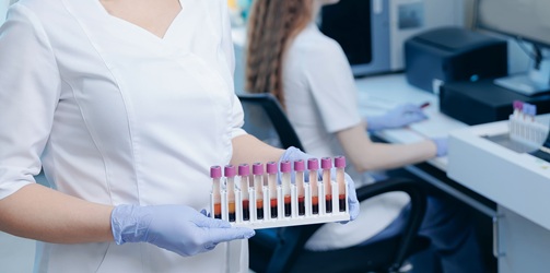
Image: The low cost microscope system constructed to perform multiple simultaneous time-lapse studies on various cell types (Photo courtesy of Adam Lynch).
Scientists at Brunel University (Uxbridge, UK) constructed the apparatus from cheaply bought materials. Various lighting sources were tested, and ultimately a light-emitting diode (LED) strip desk lamp was selected. An incubation chamber was developed to fit over the top of the stage and the chamber was made from transparent acrylic to allow visualization inside.
The three microscopes used were identical models (VMS-004D, Veho; Southampton, UK) in order to prevent any discrepancies. These microscopes use a complementary metal–oxide–semiconductor (CMOS) image sensor with 1.3 mega-pixel resolution. Magnification has two set levels, from approximately ×20 minimum to around ×400 maximum, achieved using a focusing wheel. To enhance stability, magnification and to allow for observation of live samples in liquid (cells) the microscopes were inverted.
The imaging capability of the system was compared to a conventional inverted microscope fitted with a 1.3 megapixel camera. The highest magnification on the conventional microscope was greater than the constructed system, but the maximum pixel resolution of images was the same. Spatial resolution on the conventional microscope was higher and intra-cellular detail could be seen at the highest magnification that could not be distinguished in the innovative system when images were enlarged to match the size.
The authors concluded that the novel cell tracking system had the ability to perform multiple simultaneous time-lapse studies on various cell types. Due to its low-cost, portability and commercially available components they believe that this system has the potential to enable time-lapse studies by non-specialist departments, and may be a practical solution for scientists with limited financial resources. The study was published on August 14, 2014, in the journal Public Library of Science ONE.
Related Links:
Brunel University
Veho
New
Gold Member
POC Helicobacter Pylori Test Kit
Hepy Urease Test


Pipette Calibration System
Artel PCS®

CMV CLIA Diagnostic
CLIA CMV IgA Screen Group