Smartphone Optical Sensor Diagnoses Kaposi's Sarcoma
By LabMedica International staff writers
Posted on 27 Jun 2013
A smartphone-based system has been created for in-the-field detection of the herpes virus that causes Kaposi's sarcoma, a type of cancer linked to Acquired Immunodeficiency Syndrome (AIDS).Posted on 27 Jun 2013
The system consists of a plug-in optical accessory and disposable microfluidic chips, and the technique could also be adapted for use in detecting a range of other conditions, from Escherichia coli infections to hepatitis.

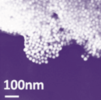
Image: Target viral DNA aggregates nanoparticles (Photo courtesy of Matthew Mancuso).
Biomedical engineers at Cornell University (Ithaca, NY, USA) devised and used the smartphones for diagnostic testing, which unlike other methods, this system is chemically based and does not use the phone's built-in camera. Instead, gold nanoparticles are combined or conjugated with short DNA snippets that bind to Kaposi's DNA sequences, and a solution with the combined particles is added to a microfluidic chip.
In the presence of viral DNA, the particles clump together, which affects the transmission of light through the solution. This causes a color change that can be measured with an optical sensor connected to a smartphone via a micro-USB (universal serial bus) port. The nanoparticle solution is a bright red when little or no Kaposi's virus DNA is present, but at higher concentrations, the solution turns a duller purple, providing a quick method to quantify the amount of Kaposi's DNA. The main advantage of the system compared to previous Kaposi's detection methods is that users can diagnose the condition with little training.
David Erickson, PhD, who devised the technique said, “The accessory provides an ultraportable way to determine whether or not viral DNA is present in a sample.” Matthew Mancuso, a doctoral student in Prof. Erickson’s laboratory added, “The smartphone reader could also work with other color-changing reactions, such as the popular enzyme-linked immunosorbent assays (ELISA), a common tool in medicine to test for human immunodeficiency virus (HIV), hepatitis, food allergens, and E. coli. The laboratory has also created smartphone accessories for use with the color-changing strips in pH and urine assays. These accessories could form the basis of a simple, at-home, personal biofluid health monitor.” The study was presented at the Conference on Lasers and Electro Optics held June 9-14, 2013, in San Jose (CA, USA).
Related Links:
Cornell University


